

Reproductive System, Female
Clitoral Gland, Duct - Dilation
Narrative
Clitoral gland duct dilation (Figure 1, Figure 2, Figure 3, Figure 4, Figure 5, Figure 6, Figure 7, Figure 8, Figure 9, Figure 10 and Figure 11) is a common incidental finding in aging rodents. It has also been induced with administration of androgenic steroids. Dilation of the ducts of the clitoral glands is frequently accompanied by atrophy of the glandular tissue. The epithelial lining of smaller ducts may be cuboidal, flattened, or keratinized squamous epithelium. When this condition is severe, the dilated ducts may occupy the majority of the clitoral gland, with remnants of the acinar tissue evident between the dilated ducts, and the glands may be grossly enlarged due to duct dilation. Care must be taken in diagnosing this lesion in mice because mice typically have very cavernous ducts with little glandular tissue.
Brix AE, Nyska A, Haseman JK, Sells DM, Jokinen MP, Walker NJ. 2005. Incidences of selected lesions in control female Harlan Sprague-Dawley rats from two-year studies performed by the National Toxicology Program. Toxicol Pathol 33:477-483.
Abstract: https://www.ncbi.nlm.nih.gov/pubmed/16036865Copeland-Haines D, Eustis SL. 1990. Specialized sebaceous glands. In: Pathology of the Fischer Rat: Reference and Atlas (Boorman GA, Eustis SL, Elwell MR, Montgomery CA, MacKenzie WF, eds). Academic Press, San Diego, CA, 279-293.
National Toxicology Program. 1999. NTP TR-485. Toxicology and Carcinogenesis Studies of Oxymetholone (CAS No. 434-07-1) in F344/N Rats and Toxicology Studies of Oxymetholone in B6C3F1 Mice (Gavage Studies). NTP, Research Triangle Park, NC.
Abstract: https://ntp.niehs.nih.gov/go/9771National Toxicology Program. 2006. NTP TR-521. Toxicology and Carcinogenesis Studies of 2,3,7,8-Tetrachlorodibenzo-p-dioxin (TCDD) (CAS No. 1746-01-6) in Female Harlan Sprague-Dawley Rats (Gavage Studies). NTP, Research Triangle Park, NC.
Abstract: https://ntp.niehs.nih.gov/go/9303National Toxicology Program. 2010. NTP TR-558. Toxicology and Carcinogenesis Studies of 3,3',4,4'-Tetrachloroazobenzene (TCAB) (CAS No. 14047-09-7) in Harlan Sprague-Dawley Rats and B6C3F1 Mice (Gavage Studies). NTP, Research Triangle Park, NC.
Abstract: https://ntp.niehs.nih.gov/go/33564Seely JC, Boorman GA. 1991. Mammary gland and specialized sebaceous glands. In: Pathology of the Mouse: Reference and Atlas (Maronpot RR, Boorman GA, Gaul BW, eds). Cache River Press, Vienna, IL, 613-635.
Clitoral gland, Duct - Dilation, Marked in a female Sprague-Dawley rat from a chronic study. The central duct is markedly dilated and filled with eosinophilic material.
All Images

Clitoral gland, Duct - Dilation, Marked in a female Sprague-Dawley rat from a chronic study. The central duct is markedly dilated and filled with eosinophilic material.

Clitoral gland, Duct - Dilation, Marked in a female Sprague-Dawley rat from a chronic study (higher magnification of Figure 1). Squamous epithelium lines the dilated duct, and eosinophilic debris is evident in the lumen.

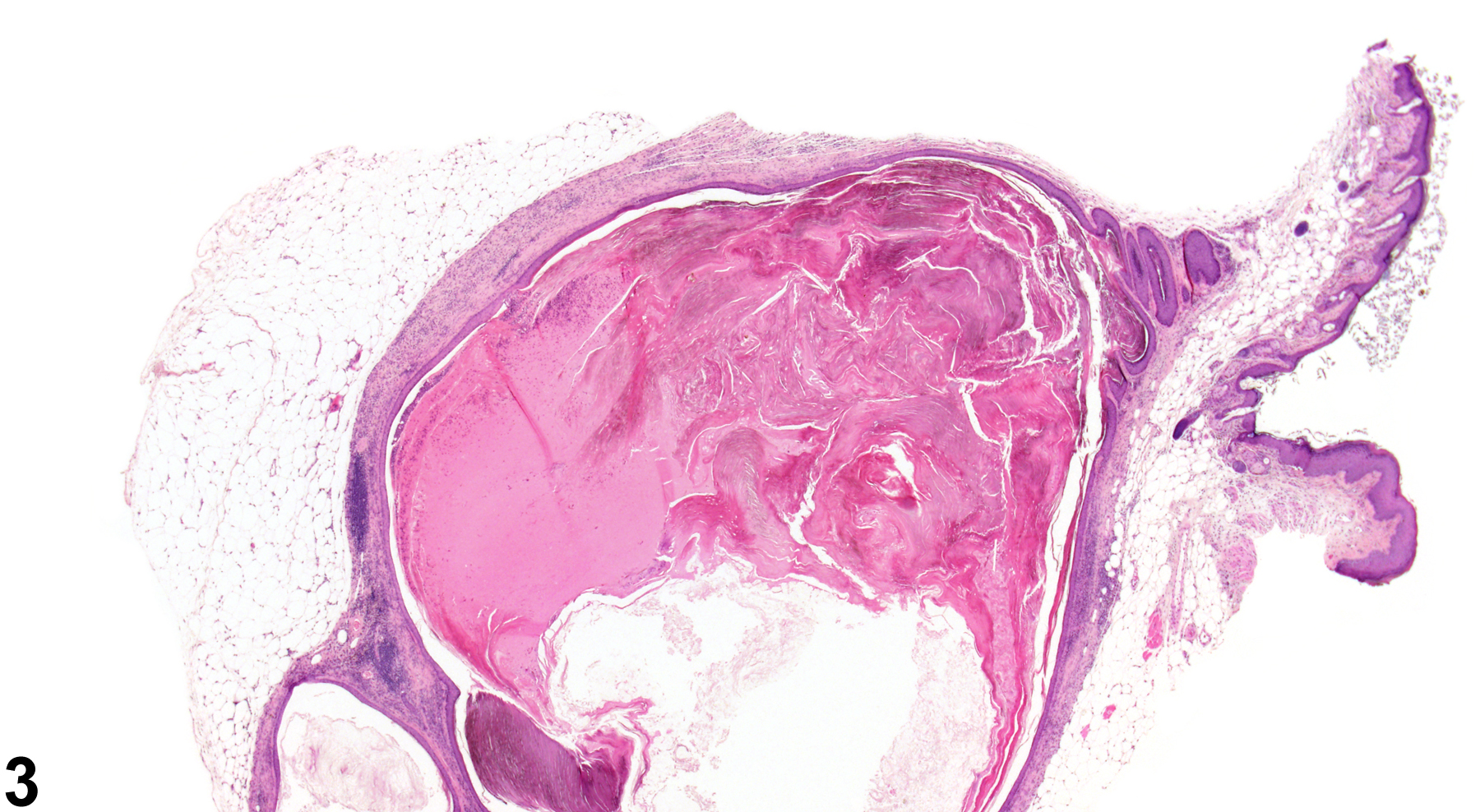
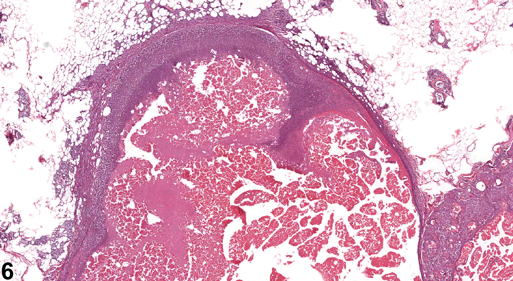
Clitoral gland, Duct - Dilation, Marked in a female B6C3F1/N mouse from a chronic study. The markedly dilated central duct is filled with eosinophilic material and occupies much of the clitoral gland.

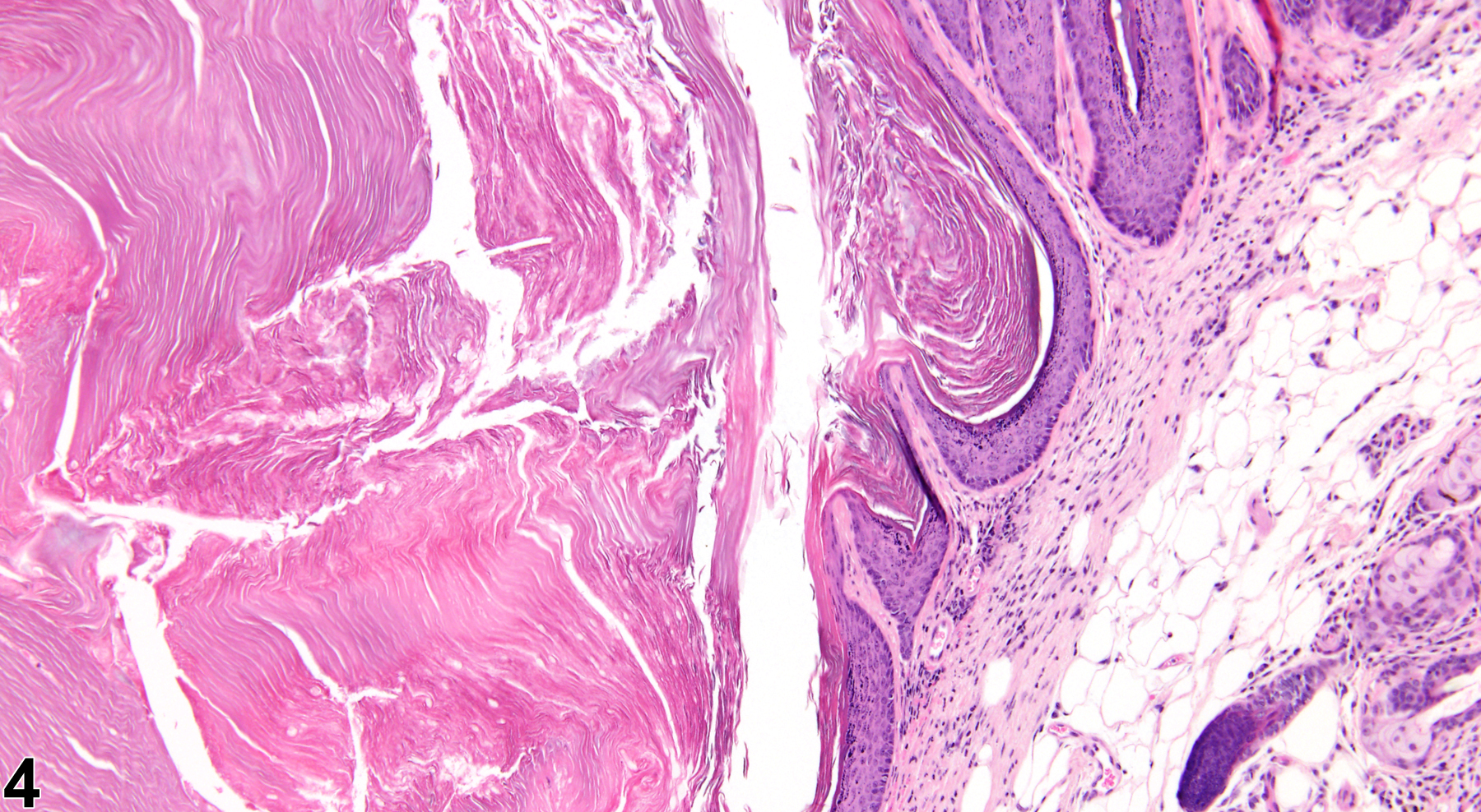
Clitoral gland, Duct - Dilation, Marked in a female B6C3F1/N mouse from a chronic study (higher magnification of Figure 3). Squamous epithelium lines the dilated duct, and eosinophilic debris is evident centrally in the lumen.

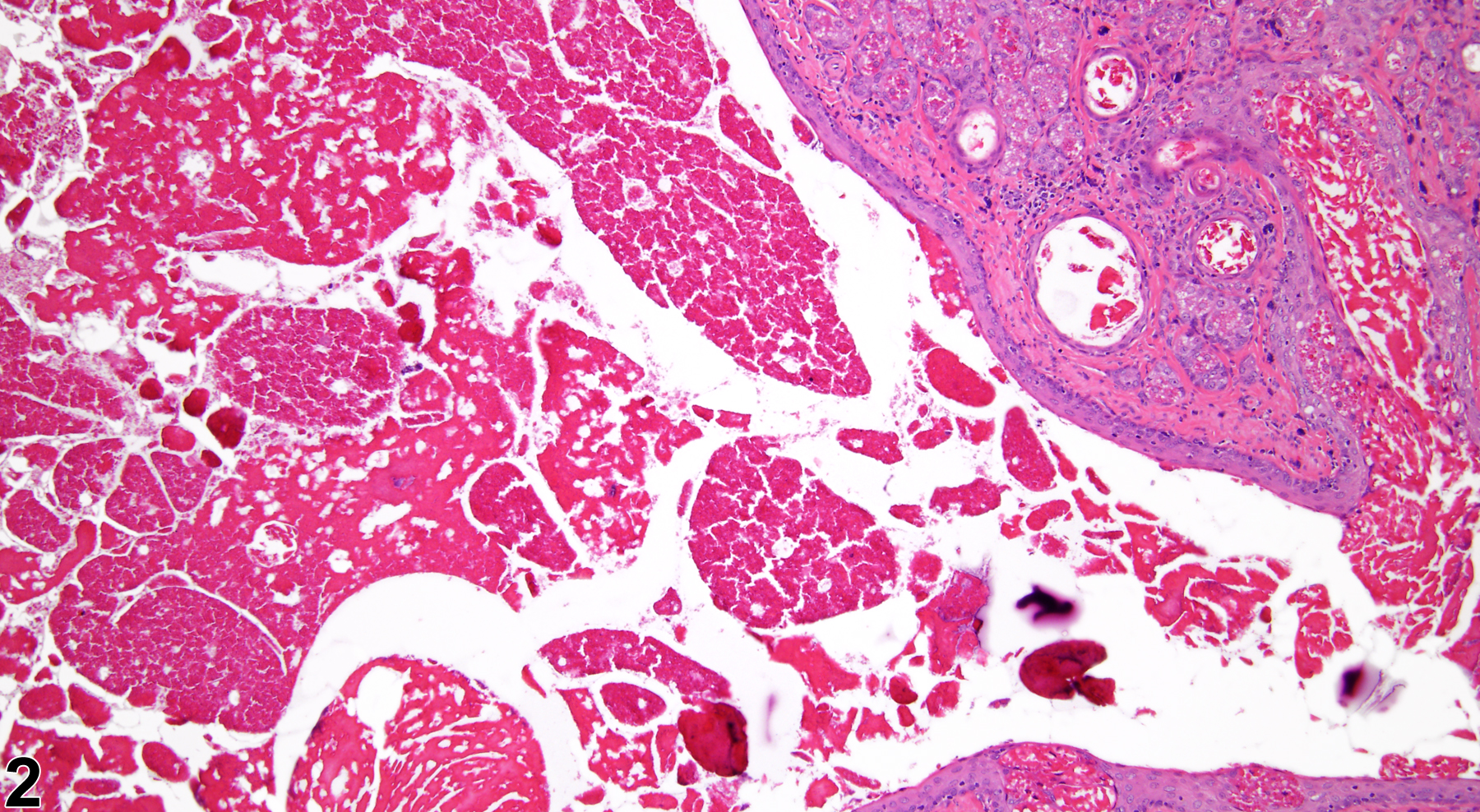
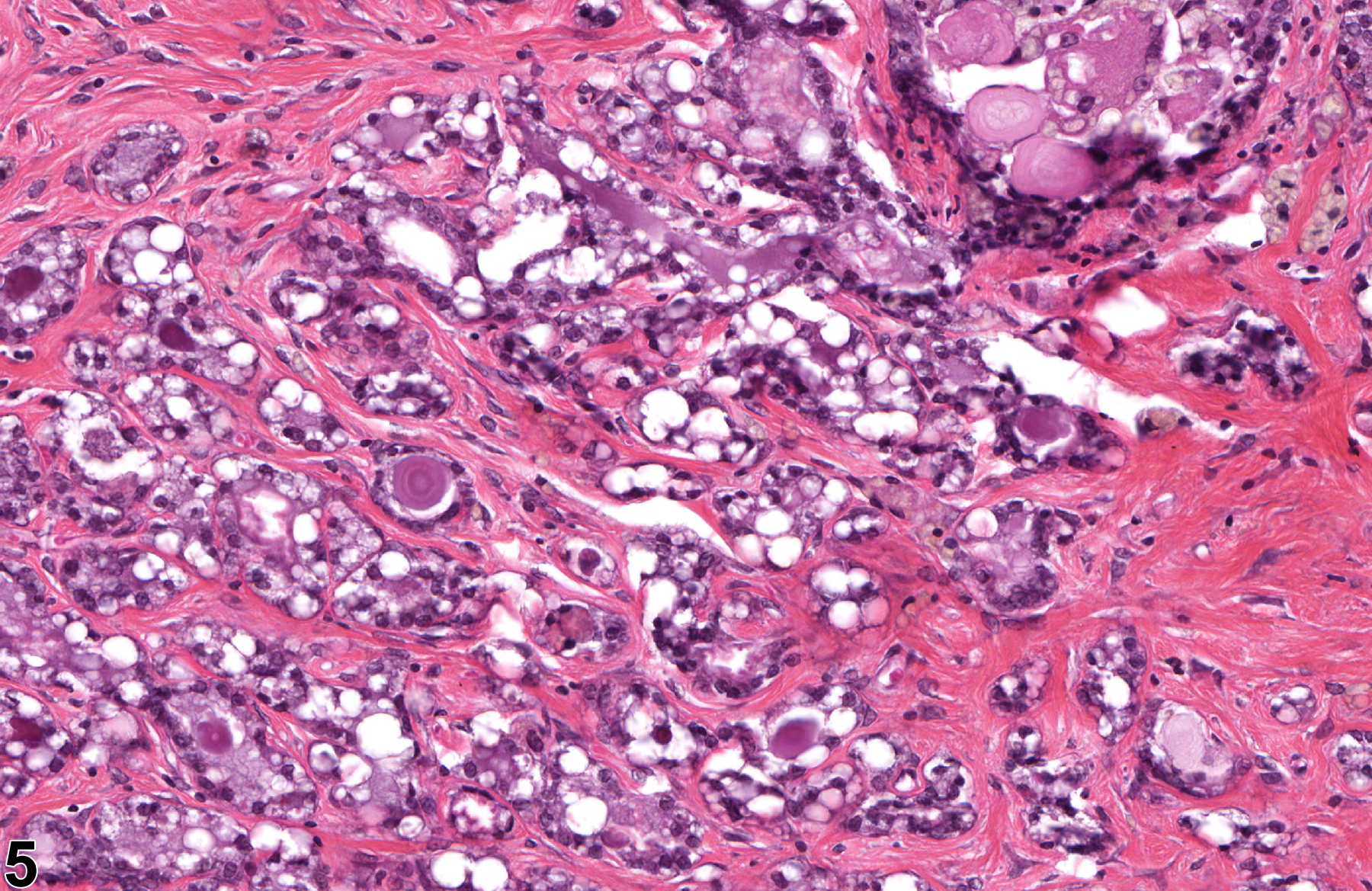
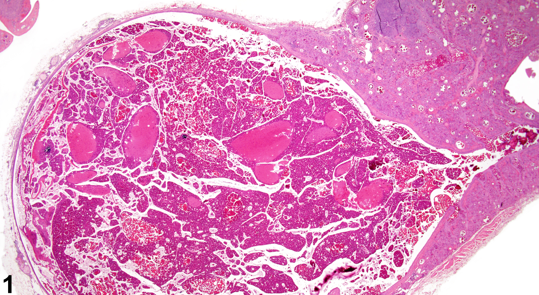
Clitoral gland, Duct - Dilation, Marked in a female F344/N rat from a chronic study. The central duct is markedly dilated and contains amorphous eosinophilic material.

Clitoral gland - Atrophy in a female F344/N rat from a chronic study (higher magnification of Figure 4). There is an increase in intervening fibrous connective tissue with a reduction in glandular tissue.

Clitoral gland, Duct - Dilation, Marked in a female F344/N rat from a chronic study (higher magnification of Figure 5). The dilated duct contains amorphous eosinophilic material and is lined by squamous epithelium.

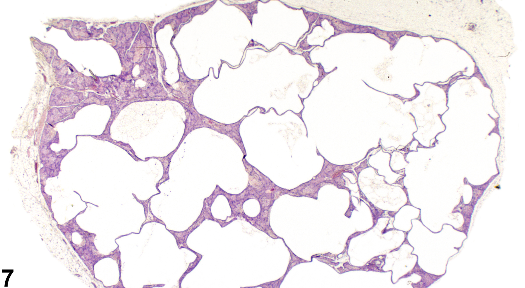
Clitoral gland, Duct - Dilation, in a female B6C3F1/N mouse from a subchronic study. Dilated ducts comprise the majority of the clitoral gland.

Clitoral gland, Duct - Dilation, Bilateral, in a female B6C3F1/N mouse from a subchronic study. Dilated ducts comprise the majority of the clitoral gland.

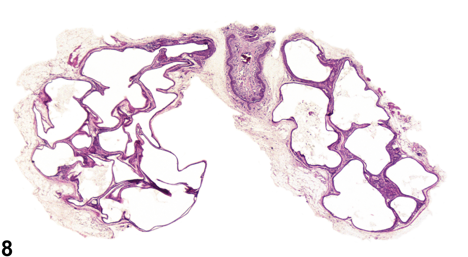
Clitoral gland, Duct - Dilation in a female B6C3F1/N mouse from a subchronic study (higher magnification of Figure 8). There are several dilated ducts and intervening acinar tissue.

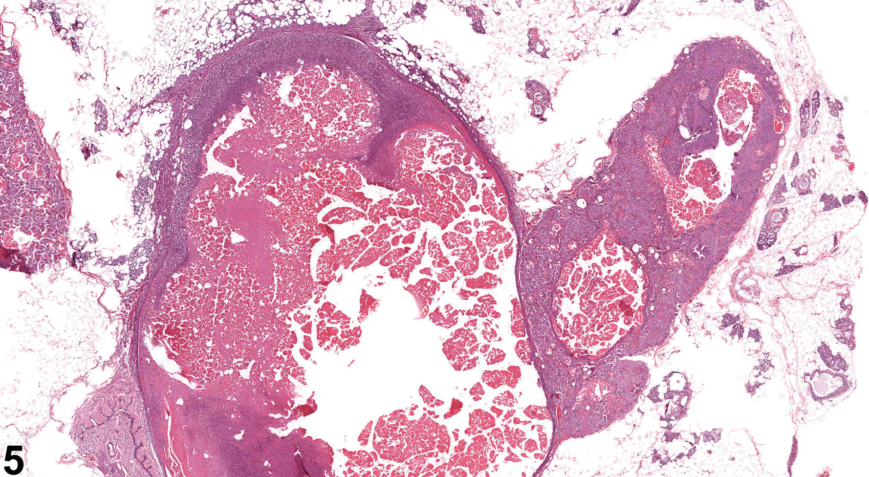
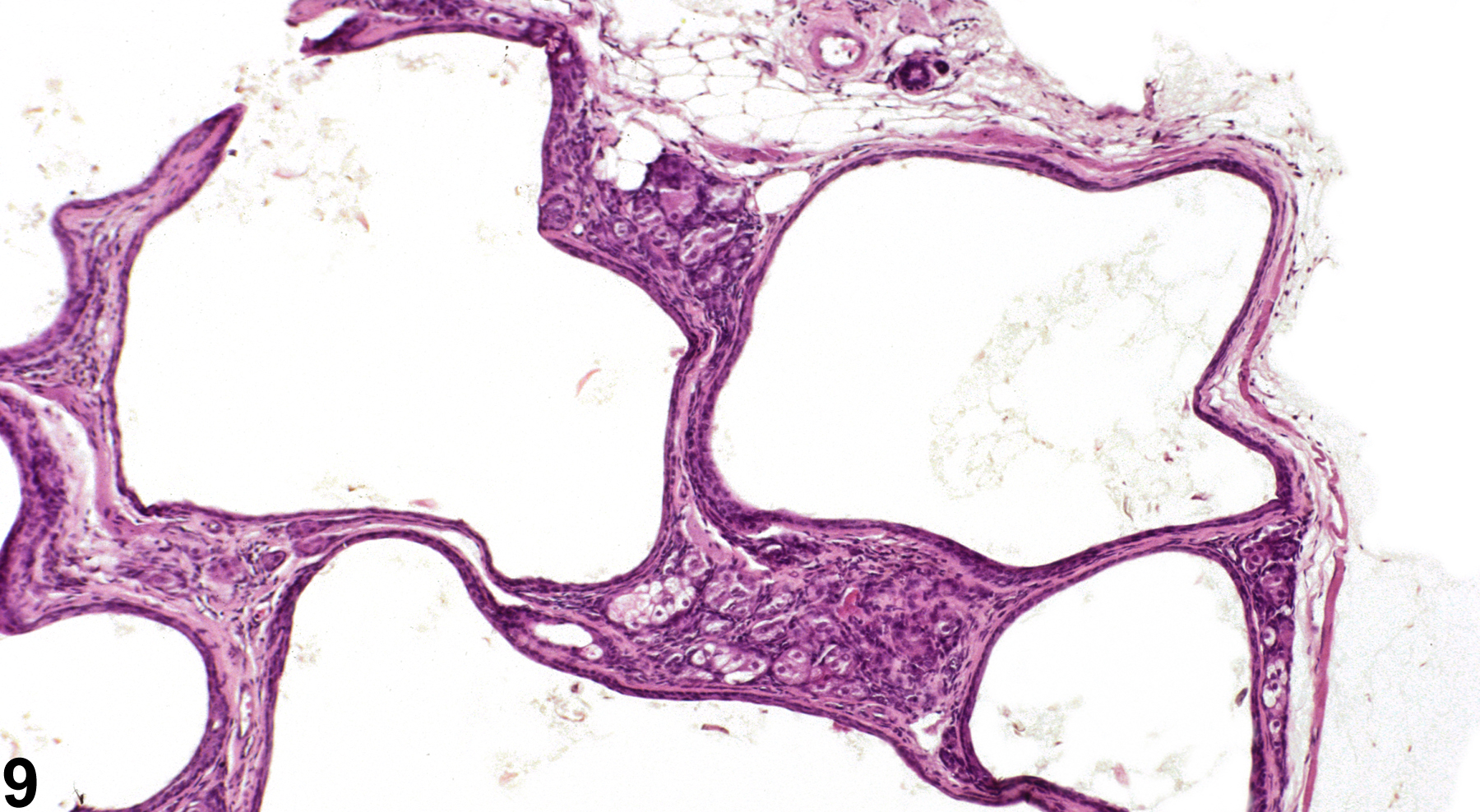
Clitoral gland, Duct - Dilation in a female F344/N rat from a subchronic study. The dilated ducts are surrounded by acinar tissue.

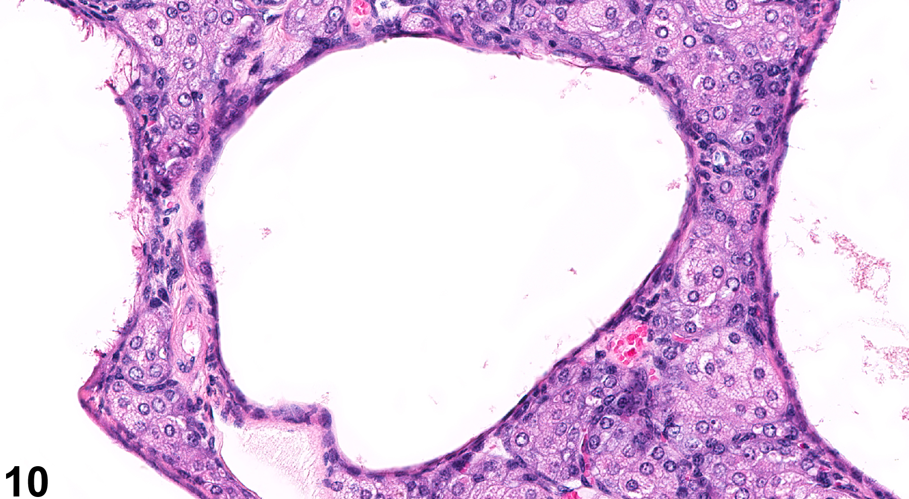
Clitoral gland, Duct - Dilation in a female B6C3F1/N mouse from a subchronic study. The ducts have a flattened epithelial lining and contain scant luminal debris.