

Reproductive System, Female
Ovary - Cyst, Epithelial
Narrative
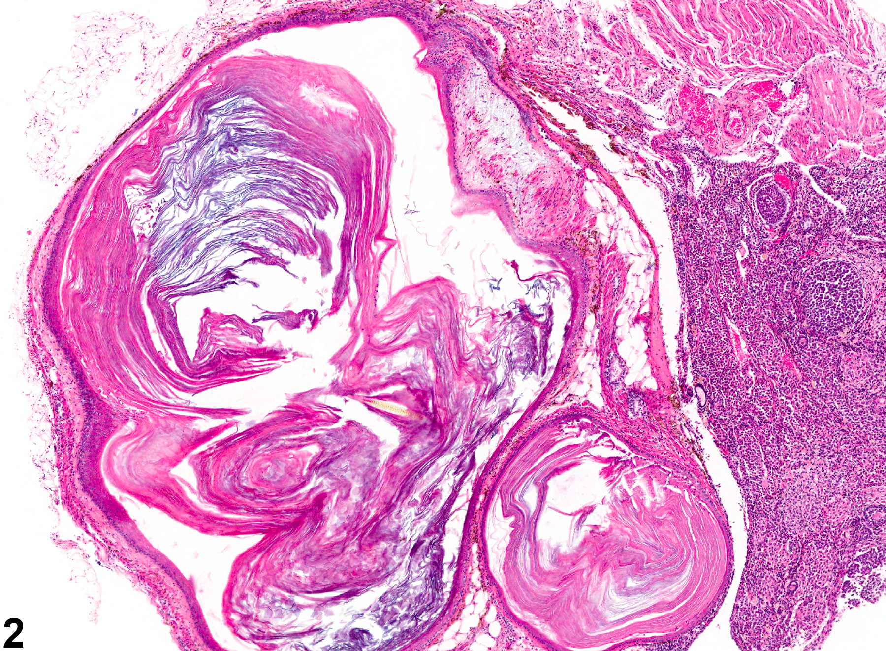
Epithelial cysts (Figure 1 and Figure 2) are seen occasionally in mice and are rarely seen in rats. These cysts are considered to arise from downgrowth of the surface epithelium of the ovary. The cysts are lined by flattened, cuboidal, columnar, or squamous epithelium; squamous epithelium may be keratinized. The lining epithelium may resemble that of the surface epithelium.
Ovarian epithelial cysts should be diagnosed, but cysts occurring as background lesions need not be graded. In the case of an epithelial cyst, the diagnosis should include the type of cyst as a modifier (i.e., Ovary - Cyst, Epithelial). If the cysts are thought to be treatment related, they may be graded to fully characterize the treatment effect. If applicable, the terms "bilateral" and "multiple" may be included in the diagnosis.
Davis BJ, Dixon D, Herbert RA. 1999. Ovary, oviduct, uterus, cervix and vagina. In: Pathology of the Mouse: Reference and Atlas (Maronpot RR, Boorman GA, Gaul BW, eds). Cache River Press, Vienna, IL, 409-444.
Dixon D, Alison R, Bach U, Colman K, Foley GL, Harleman JH, Hawarth R, Herbert R, Heuser A, Long G, Mirsky M, Regan K, Van Esch E, Westwood FR, Vidal J, Yoshida M. 2014. Nonproliferative and proliferative lesions of the rat and mouse female reproductive system (INHAND). J Toxicol Pathol 27(suppl):1S-107S.
Full Text: https://www.ncbi.nlm.nih.gov/pmc/articles/PMC4253081/Faccini JM, Abbott DP, Paulus GJJ. 1990. Female genital tract. In: Mouse Histopathology: A Glossary for Use in Toxicity and Carcinogenicity Studies (Greaves P, Faccini JM, eds). Elsevier, Amsterdam, 147-168.
Montgomery CA, Alison RH. 1987. Non-neoplastic lesions of the ovary in Fischer 344 rats and B6C3F1 mice. Environ Health Perspect 73:53-75.
Abstract: https://www.ncbi.nlm.nih.gov/pmc/articles/PMC1474552/National Toxicology Program. 1995. NTP TR-438. Toxicology and Carcinogenesis Studies of Benzethonium Chloride (CAS No. 121-54-0) in F344/N Rats and B6C3F1 Mice (Dermal Studies). NTP, Research Triangle Park, NC.
Abstract: https://ntp.niehs.nih.gov/go/6020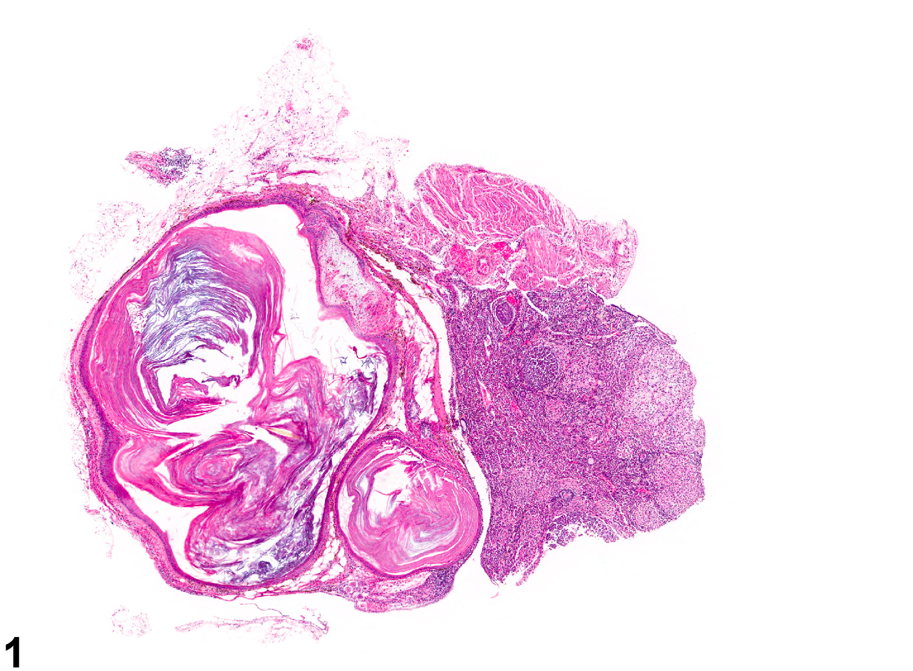
Ovary - Cyst, Epithelial in a female B6C3F1/N mouse from a chronic study. The cyst is compressing adjacent ovarian parenchyma.
All Images

Ovary - Cyst, Epithelial in a female B6C3F1/N mouse from a chronic study. The cyst is compressing adjacent ovarian parenchyma.

Ovary - Cyst, Epithelial in a female B6C3F1/N mouse from a chronic study (higher magnification of Figure 1). Two adjacent epithelial cysts are lined by stratified squamous epithelium and contain keratin.