

Reproductive System, Female
Ovary - Hyperplasia, Sertoliform
Narrative
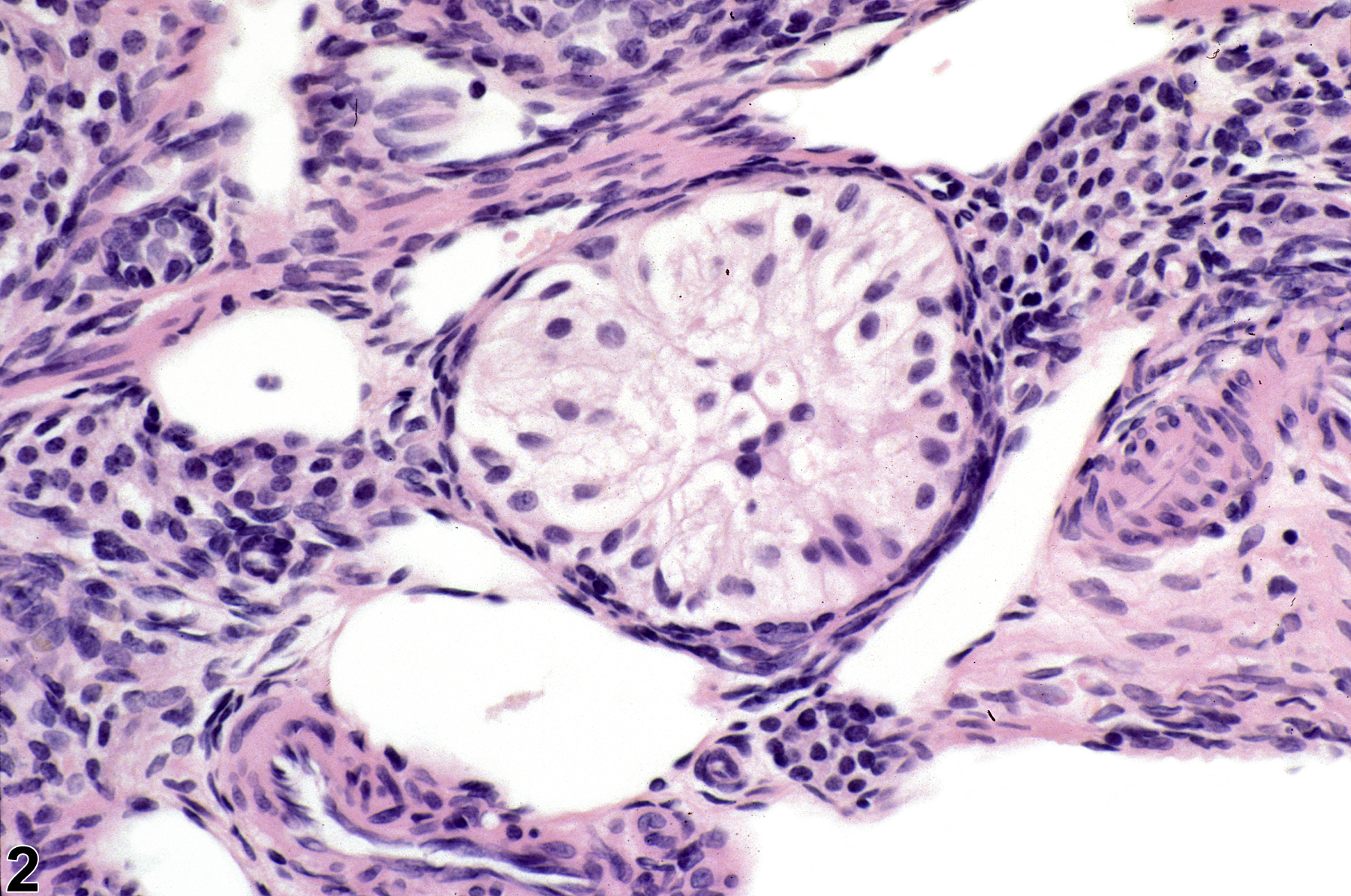
The key features of Sertoli cell hyperplasia in the ovary are that the cells resemble their testicular counterpart histologically and that these proliferative lesions often arise in the region of the hilus (Figure 1). They are characterized by seminiferous-like tubules lined by cells with basally located nuclei and abundant, faintly eosinophilic, vacuolated cytoplasm extending into the lumen (Figure 2). Sometimes there may be areas with focal nests of Sertoli cells without obvious tubular structures. Sertoli cell hyperplasia is typically a focal lesion smaller than or equal to the size of a corpus luteum; there is no cellular atypia, and neither compression nor capsules are present.
Sertoli cell hyperplasia is thought to be derived from stromal cells in atrophic ovaries. This finding is associated with aging but may also be induced by chemicals. Sertoli cell hyperplasia must be differentiated from dysgerminoma and Sertoli cell tumor. Sertoli cell tumors are larger than the size of a corpus luteum; there is compression of adjacent tissue, and some cellular atypia may be present. Dysgerminomas are not reported in the rat and are a rare tumor in mice; the histologic appearance is of sheets of large, round, undifferentiated cells.
Alison RH, Morgan KT, Montgomery CA. 1990. Ovary. In: Pathology of the Fischer Rat: Reference and Atlas (Boorman GA, Eustis SL, Elwell MR, Montgomery CA, MacKenzie WF, eds). Academic Press, San Diego, CA, 429-442.
Dixon D, Alison R, Bach U, Colman K, Foley GL, Harleman JH, Hawarth R, Herbert R, Heuser A, Long G, Mirsky M, Regan K, Van Esch E, Westwood FR, Vidal J, Yoshida M. 2014. Nonproliferative and proliferative lesions of the rat and mouse female reproductive system (INHAND). J Toxicol Pathol 27(suppl):1S-107S.
Full Text: https://www.ncbi.nlm.nih.gov/pmc/articles/PMC4253081/Gopinath C, Prentice DE, Lewis DJ. 1987. Atlas of Experimental Toxicological Pathology. MTP Press, Lancaster, UK.
Greaves P. 2012. Female genital tract. In: Histopathology of Preclinical Toxicity Studies: Interpretation and Relevance in Drug Safety Evaluation, 4th ed. Elsevier, Amsterdam, 667-724.
Maekawa A, Yoshida A. 1996. Susceptibility of the female genital system to toxic substances. In: Pathobiology of the Aging Mouse, Vol 1 (Mohr U, Dungworth DL, Capen CC, Carlton WW, Sundberg JP, Ward JM, eds). ILSI Press, Washington, DC, 481-493.
Mann PC. 1997. Selected lesions of dioxin in laboratory rodents. Toxicol Pathol 25:72-79.
Abstract: https://www.ncbi.nlm.nih.gov/pubmed/9061855Montgomery CA, Alison RH. 1987. Non-neoplastic lesions of the ovary in Fischer 344 rats and B6C3F1 mice. Environ Health Perspect 73:53-75.
Abstract: https://www.ncbi.nlm.nih.gov/pmc/articles/PMC1474552/National Toxicology Program. 1987. NTP TR-315. Toxicology and Carcinogenesis Studies of Oxytetracycline Hydrochloride (CAS No. 2058-46-0) in F344/N Rats and B6C3F1 Mice (Feed Studies). NTP, Research Triangle Park, NC.
Abstract: https://ntp.niehs.nih.gov/go/9840National Toxicology Program. 2006. NTP TR-521. Toxicology and Carcinogenesis Studies of 2,3,7,8-Tetrachlorodibenzo-p-dioxin (TCDD) (CAS No. 1746-01-6) in Female Harlan Sprague-Dawley Rats (Gavage Studies). NTP, Research Triangle Park, NC.
Abstract: https://ntp.niehs.nih.gov/go/9303Peluso JJ, Gordon LR. 1992. Nonneoplastic and neoplastic changes in the ovary. In: Pathobiology of the Aging Rat (Mohr U, Dungworth DL, Capen CC, eds). ILSI Press, Washington, DC, 351-364.
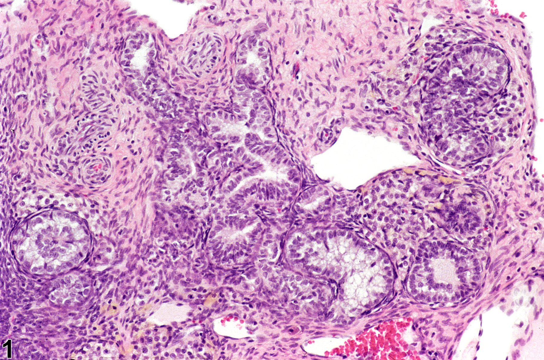
Ovary - Hyperplasia, Sertoliform in a female F344/N rat from a chronic study. The hyperplastic tubules are adjacent to the hilus of the ovary.
All Images

Ovary - Hyperplasia, Sertoliform in a female F344/N rat from a chronic study. The hyperplastic tubules are adjacent to the hilus of the ovary.

Ovary - Hyperplasia, Sertoliform in a female F344/N rat from a chronic study (higher magnification of Figure 1). The tubules are lined by columnar cells with indistinct cytoplasmic borders and basal nuclei.