

Reproductive System, Female
Ovary, Interstitial Cell - Hyperplasia
Narrative
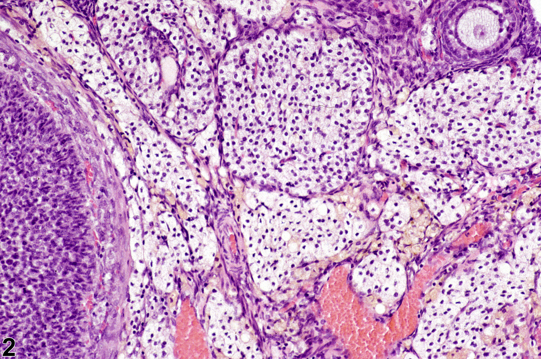
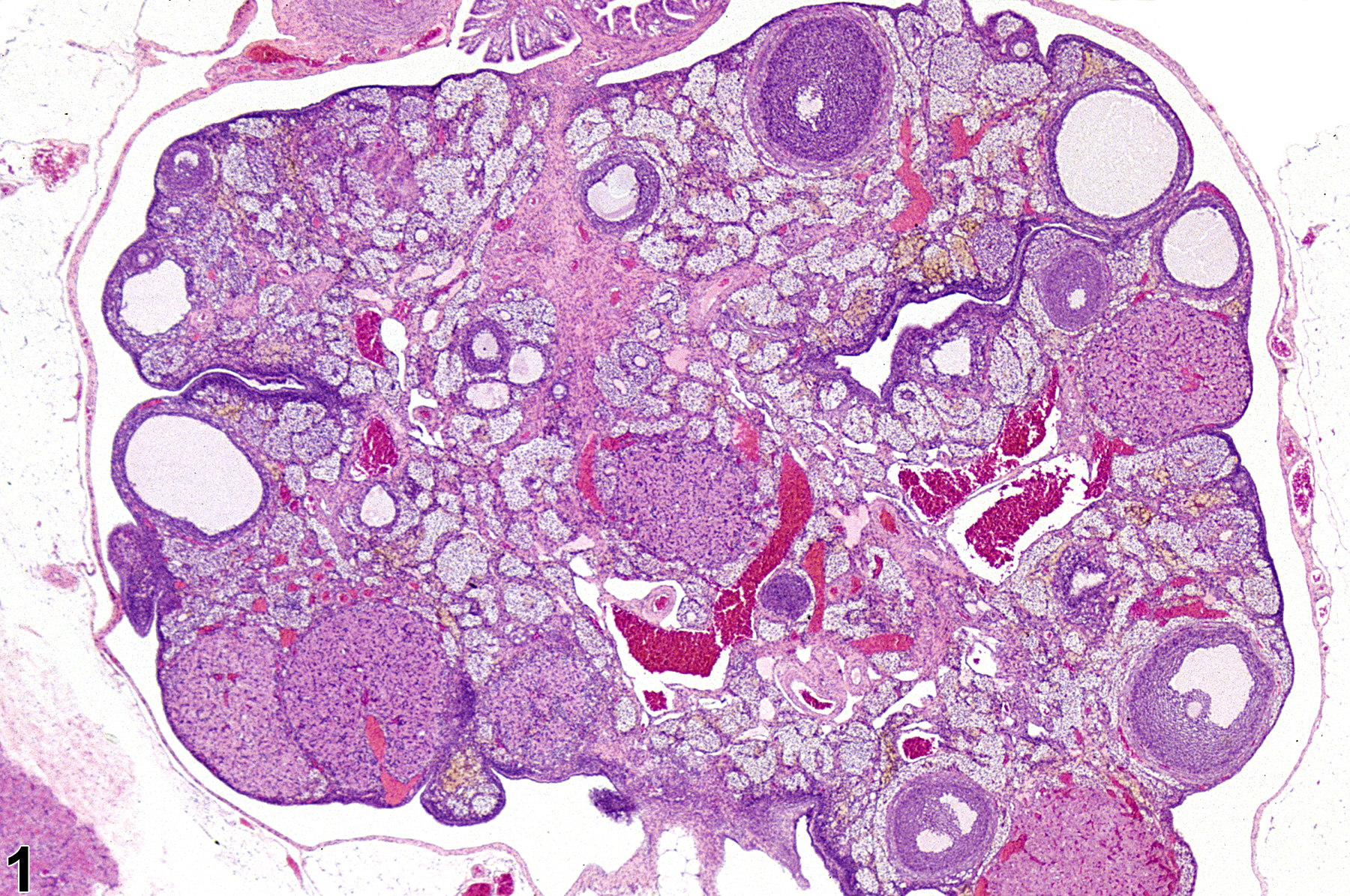
Typically in interstitial cell hyperplasia many variably sized clusters of pale-staining cells are interspersed among several follicles in various stages of development, including large atretic follicles, mature corpora lutea, areas of pigment deposition, and congested blood vessels (Figure 1). Cords and nests of hyperplastic interstitial cells are composed of large polyhedral cells with pale eosinophilic granular cytoplasm and central nuclei (Figure 2). Ovarian interstitial cell hyperplasia is a nonneoplastic proliferation of cellular components of the interstitial gland, derived from atretic follicles, corpora lutea, or ovarian stromal cells. This is a common aging change in rats and mice. It may also be induced by treatment with certain chemicals, such as gonadotropins and some organophosphate compounds.
Ovary, Interstitial cell - Hyperplasia should be diagnosed and graded whenever present.
Alison RH, Morgan KT, Montgomery CA. 1990. Ovary. In: Pathology of the Fischer Rat: Reference and Atlas (Boorman GA, Eustis SL, Elwell MR, Montgomery CA, MacKenzie WF, eds). Academic Press, San Diego, CA, 429-442.
Davis BJ, Dixon D, Herbert RA. 1999. Ovary, oviduct, uterus, cervix and vagina. In: Pathology of the Mouse: Reference and Atlas (Maronpot RR, Boorman GA, Gaul BW, eds). Cache River Press, Vienna, IL, 409-444.
Dixon D, Alison R, Bach U, Colman K, Foley GL, Harleman JH, Hawarth R, Herbert R, Heuser A, Long G, Mirsky M, Regan K, Van Esch E, Westwood FR, Vidal J, Yoshida M. 2014. Nonproliferative and proliferative lesions of the rat and mouse female reproductive system (INHAND). J Toxicol Pathol 27(suppl):1S-107S.
Full Text: https://www.ncbi.nlm.nih.gov/pmc/articles/PMC4253081/Greaves P. 2012. Female genital tract. In: Histopathology of Preclinical Toxicity Studies: Interpretation and Relevance in Drug Safety Evaluation, 4th ed. Elsevier, Amsterdam, 667-724.
National Toxicology Program. 1994. NTP TR-433. Toxicology and Carcinogenesis Studies of Tricresyl Phosphate (CAS No. 1330-78-5) in F344/N Rats and B6C3F1 Mice (Gavage and Feed Studies). NTP, Research Triangle Park, NC.
Abstract: https://ntp.niehs.nih.gov/go/6010Peluso JJ, Gordon LR. 1992. Nonneoplastic and neoplastic changes in the ovary. In: Pathobiology of the Aging Rat (Mohr U, Dungworth DL, Capen CC, eds). ILSI Press, Washington, DC, 351-364.
Ovary, Interstitial cell - Hyperplasia in a female F344/N rat from a subchronic study. Clusters of pale interstitial cells are interspersed among follicles.
All Images

Ovary, Interstitial cell - Hyperplasia in a female F344/N rat from a subchronic study. Clusters of pale interstitial cells are interspersed among follicles.

Ovary, Interstitial cell - Hyperplasia in a female F344/N rat from a subchronic study (higher magnification of Figure 1). There are cords and nests of hyperplastic interstitial cells, which are composed of large polyhedral cells with pale eosinophilic granular cytoplasm and a central nucleus.