

Reproductive System, Female
Uterus - Hyperplasia, Atypical
Narrative
National Toxicology Program. 2014. NTP TR-587. Toxicology Studies of Tetrabromobisphenol A (CAS No. 79-94-7) in F344/NTac Rats and B6C3F1/N Mice and Toxicology and Carcinogenesis Studies of Tetrabromobisphenol A in Wistar Han [Crl:WI(Han)] Rats and B6C3F1 Mice (Gavage Studies). NTP, Research Triangle Park, NC.
Abstract: https://ntp.niehs.nih.gov/go/41452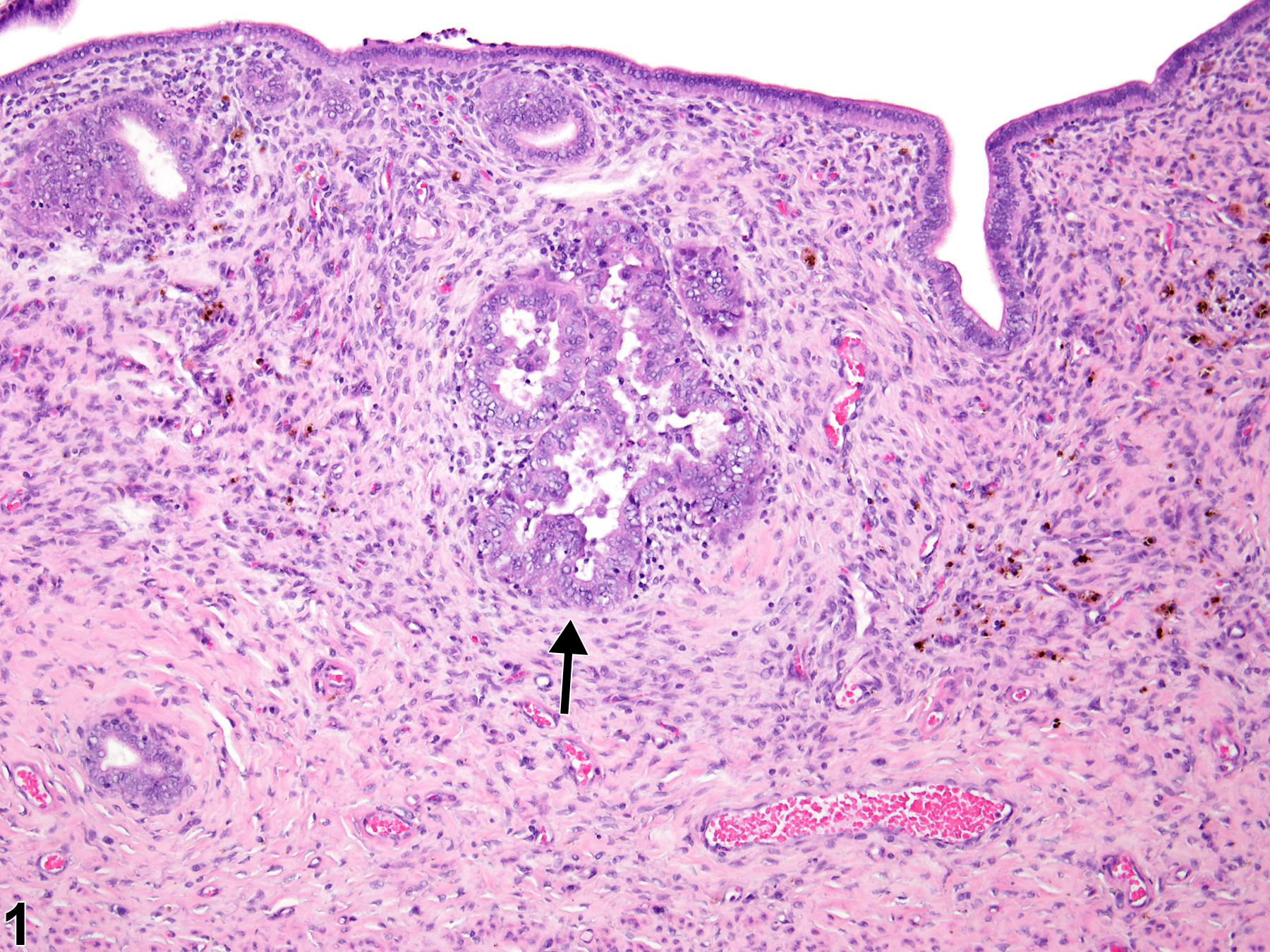
Uterus, Endometrium - Hyperplasia, Atypical in a female Wistar Han rat from a chronic study. The glandular epithelium projects into the glandular lumen (arrow), forming multiple thickened infoldings and projections.
All Images

Uterus, Endometrium - Hyperplasia, Atypical in a female Wistar Han rat from a chronic study. The glandular epithelium projects into the glandular lumen (arrow), forming multiple thickened infoldings and projections.

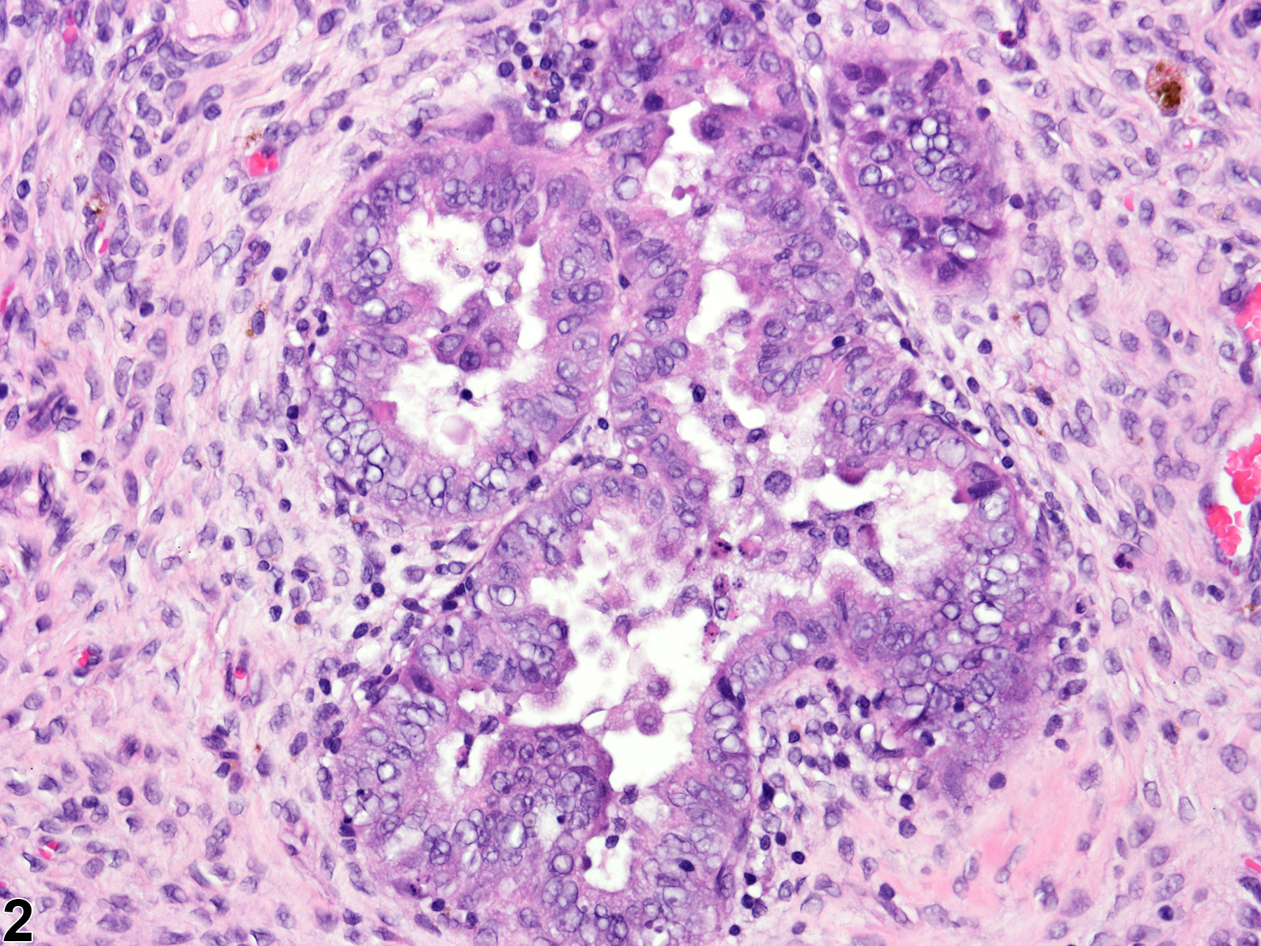
Uterus, Endometrium - Hyperplasia, Atypical in a female Wistar Han rat from a chronic study (higher magnification of Figure 1). There are multiple infoldings and projections of the endometrium into the uterine lumen.

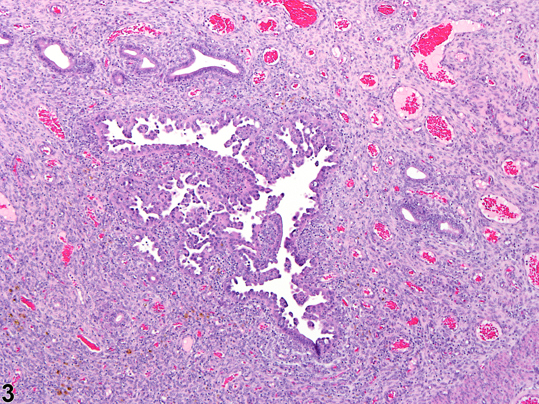
Uterus, Endometrium - Hyperplasia, Atypical in a female Wistar Han rat from a chronic study. The epithelium lining the uterine lumen projects into the lumen in irregular papillary fronds.

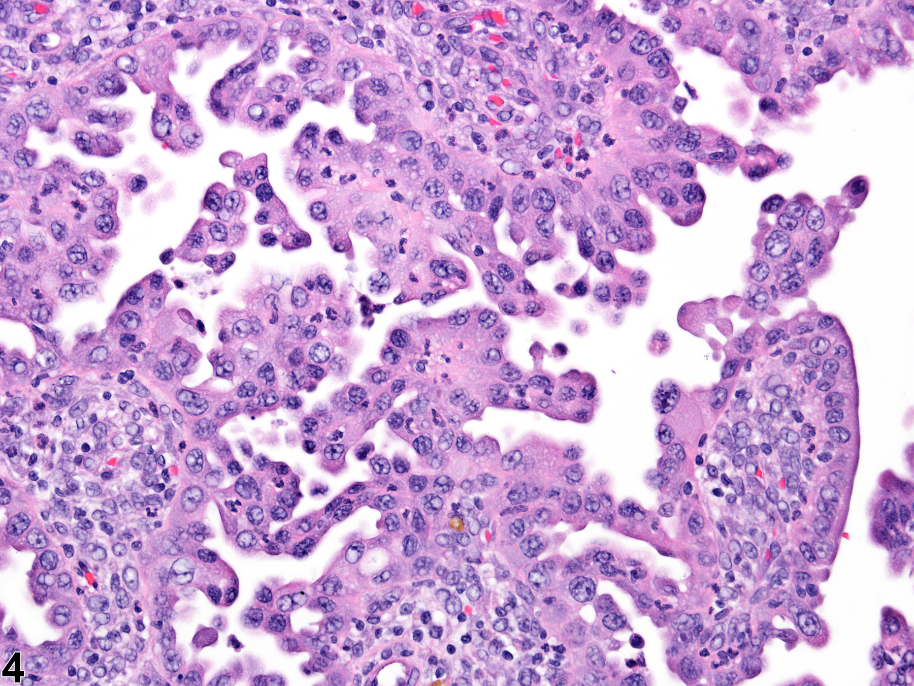
Uterus, Endometrium - Hyperplasia, Atypical in a female Wistar Han rat from a chronic study (higher magnification of Figure 3). The epithelial cells are irregularly arranged and exhibit cellular pleomorphism.

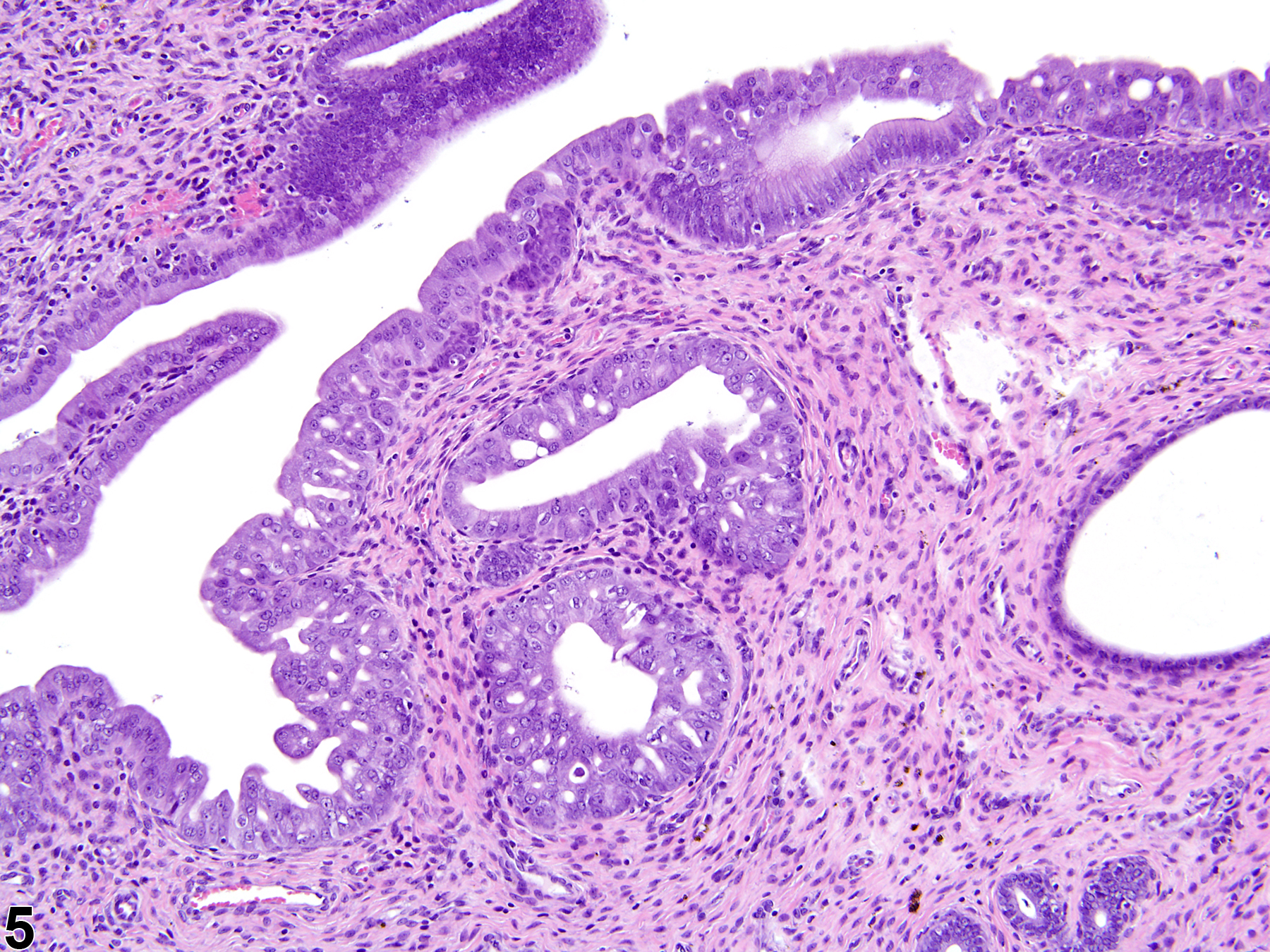
Uterus - Hyperplasia, Atypical in a female Harlan Sprague-Dawley rat from a chronic study. There is atypical hyperplasia of both surface and glandular epithelium.