

Reproductive System, Female
Uterus - Necrosis
Narrative
Uterus - Necrosis should be diagnosed and graded. If the necrosis is secondary to another lesion, such as inflammation, it should not be diagnosed separately unless warranted by severity. Lesions secondary to necrosis, such as inflammation or hemorrhage, should not be diagnosed separately unless warranted by severity. The pathologist should note in the pathology narrative if the necrosis is thought to represent an infarct.
National Toxicology Program. 1990. NTP TR-393. Toxicology and Carcinogenesis Studies of Sodium Fluoride (CAS No. 7681-49-4) in F344/N Rats and B6C3F1 Mice (Drinking Water Studies). NTP, Research Triangle Park, NC.
Abstract: https://ntp.niehs.nih.gov/go/12237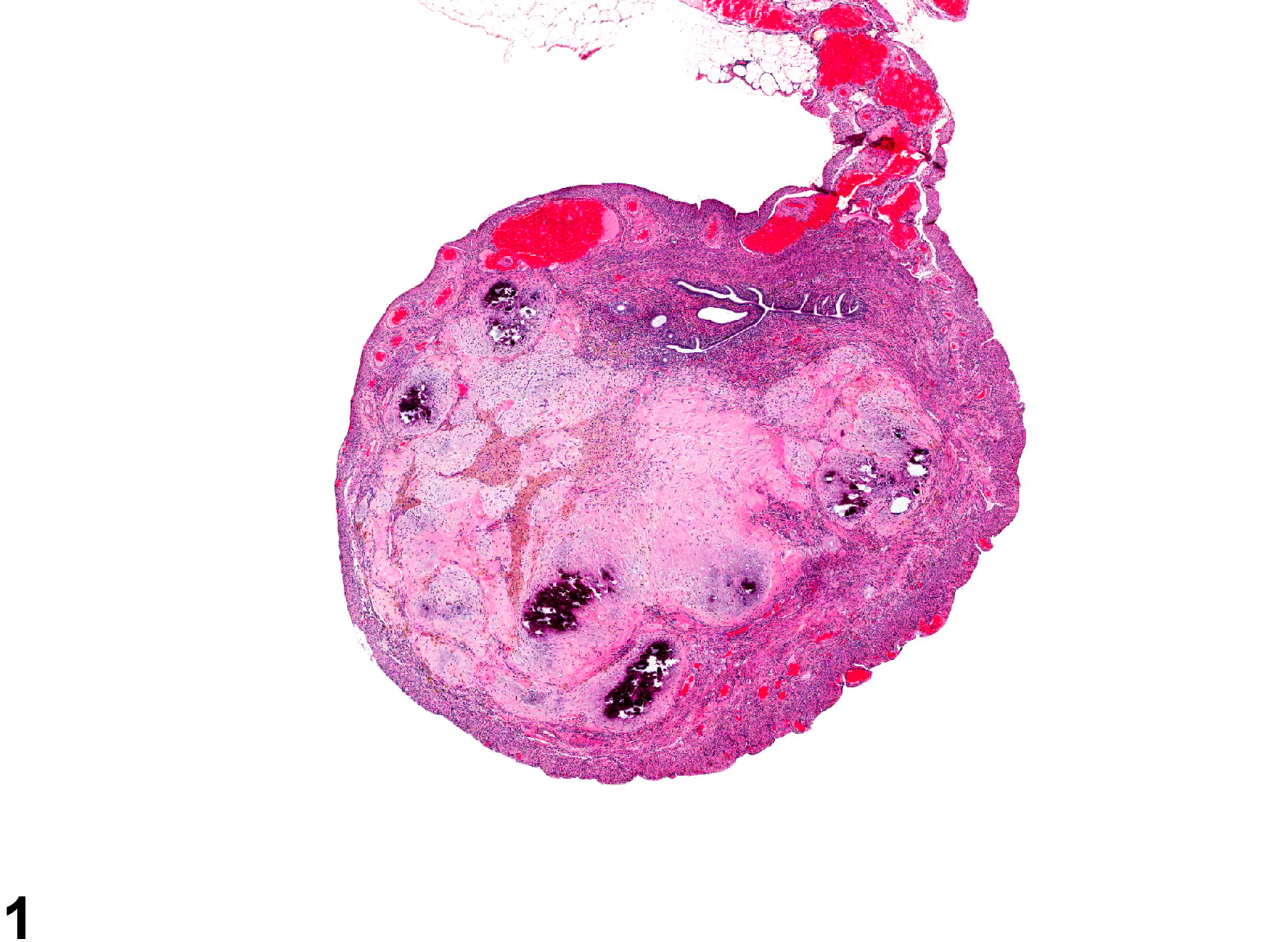
Uterus - Necrosis in a female F344/N rat from a chronic study. Fibrosis and mineralization are present in the necrotic uterine tissue.
All Images

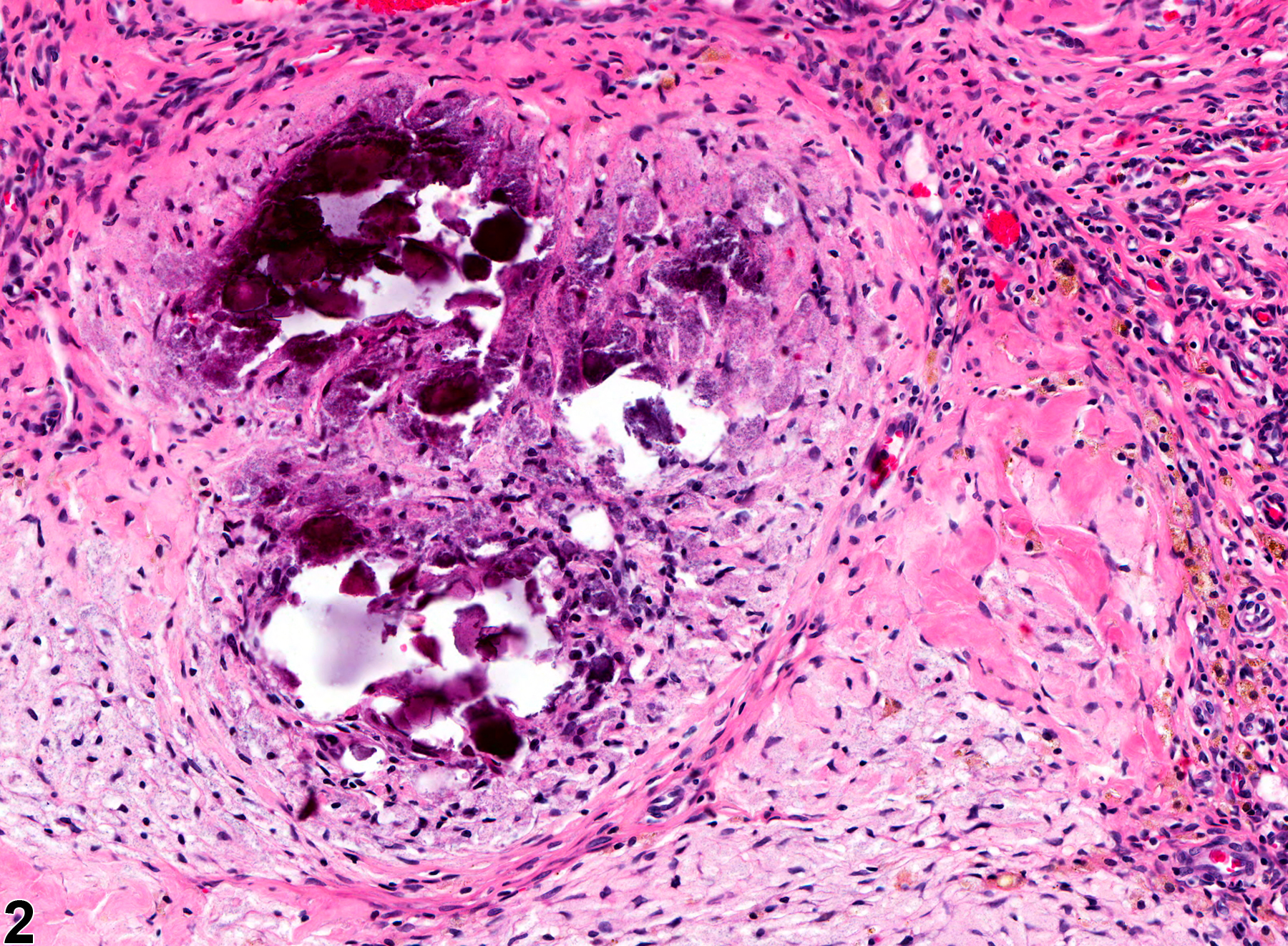
Uterus - Necrosis in a female F344/N rat from a chronic study. Fibrosis and mineralization are present in the necrotic uterine tissue.

Uterus - Necrosis in a female F344/N rat from a chronic study (higher magnification of Figure 1). There is fibrosis and mineralization of the necrotic uterine tissue.