

Reproductive System, Female
Vagina, Epithelium - Hyperplasia
Narrative
Hyperplasia of the rodent vaginal epithelium (Figure 1, Figure 2, Figure 3 and Figure 4) is often associated with hyperkeratosis and is usually diffuse. Cells are well differentiated with no atypia or invasion of the underlying stroma. Hyperplastic cells may form downward projections into the underlying stroma (rete pegs) (Figure 3 and Figure 4). Hyperplasia occurs in a variety of settings, including control rats in persistent estrus during reproductive senescence, and secondary to increases in endogenous estradiol production or administration of hormonally active xenobiotics. Focal hyperplasia of vaginal epithelium is uncommon but has been observed.
Hyperplasia should be differentiated from normal variation in the appearance of the vagina during the estrus cycle (see Introduction for description of cyclical changes). Hyperplasia is increased thickness of the vaginal epithelium (compared with normal estrus in a normally cycling animal).
The differential diagnoses for focal hyperplasia are papilloma and polyp. Hyperplastic cells are evident above the basement membrane of the epithelium and do not form a discrete proliferative mass of epithelial cells. Hyperplasia is restricted to the normal mucosal surface and does not protrude into the lumen. A polyp is composed predominantly of a fibromuscular core covered by normal or slightly hyperkeratinized epithelium.
Greaves P. 2012. Female genital tract. In: Histopathology of Preclinical Toxicity Studies: Interpretation and Relevance in Drug Safety Evaluations, 4th ed. Elsevier, Amsterdam, 667-724.
National Toxicology Program. 2010. NTP TR-558. Toxicology and Carcinogenesis Studies of 3,3',4,4'-Tetrachloroazobenzene (TCAB) (CAS No. 14047-09-7) in Harlan Sprague-Dawley Rats and B6C3F1 Mice (Gavage Studies). NTP, Research Triangle Park, NC.
Abstract: https://ntp.niehs.nih.gov/go/33564Westwood FR. 2008. The female rat reproductive cycle: A practical histological guide to staging. Toxicol Pathol 36:375-384.
Abstract: https://www.ncbi.nlm.nih.gov/pubmed/18441260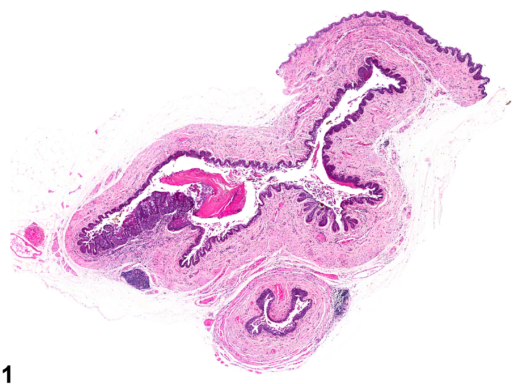
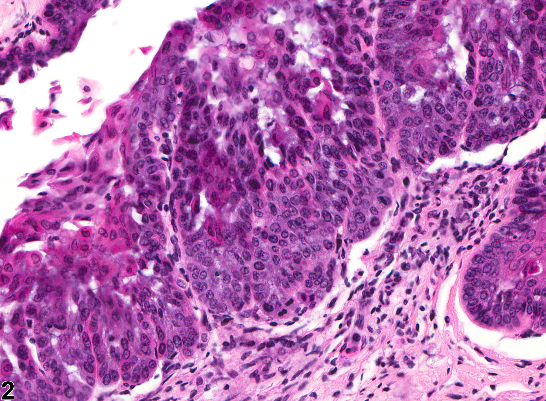
Vagina, Epithelium - Hyperplasia in a female B6C3F1/N mouse from a chronic study. There is a focal area of basal cell hyperplasia.
All Images

Vagina, Epithelium - Hyperplasia in a female B6C3F1/N mouse from a chronic study. There is a focal area of basal cell hyperplasia.

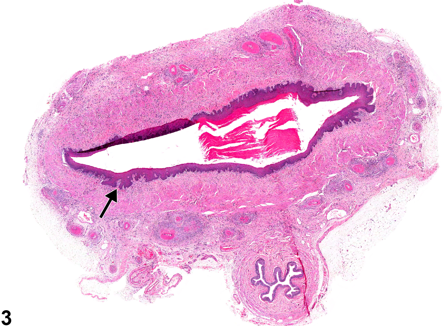
Vagina, Epithelium - Hyperplasia in a female B6C3F1/N mouse from a chronic study (higher magnification of Figure 1). There is a focal area of basal cell hyperplasia.

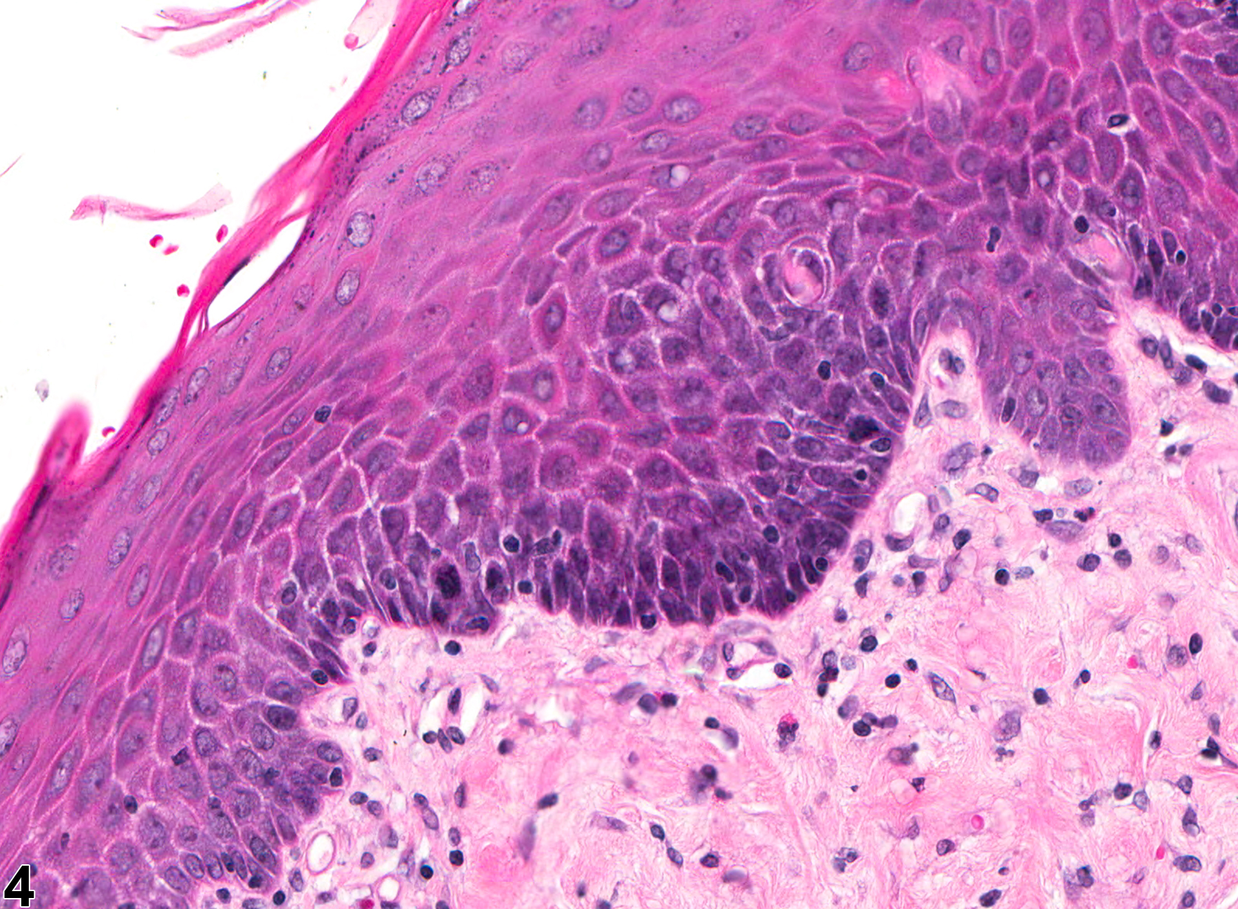
Vagina, Epithelium - Hyperplasia in a female Sprague-Dawley rat from a chronic study. Arrow indicates focal area of squamous hyperplasia.

Vagina, Epithelium - Hyperplasia in a female Sprague-Dawley rat from a chronic study (higher magnification of Figure 3). There is an area of squamous hyperplasia.