

Hepatobiliary System
Gallbladder, Epithelium - Hyperplasia
Narrative
Greaves P. 2007. Histopathology of Preclinical Toxicity Studies: Interpretation and Relevance in Drug Safety Evaluation, 3rd ed. Elsevier, Amsterdam.
Abstract: http://www.sciencedirect.com/science/book/9780444527714Harada T, Enomoto A, Boorman GA, Maronpot RR. 1999. Liver and gallbladder. In: Pathology of the Mouse: Reference and Atlas (Maronpot RR, Boorman GA, Gaul BW, eds). Cache River Press, Vienna, IL, 119-183.
Thoolen B, Maronpot RR, Harada T, Nyska A, Rousseaux C, Nolte T, Malarkey D, Kaufmann W, Kutter K, Deschl U, Nakae D, Gregson R, Winlove M, Brix A, Singl B, Belpoggi F, Ward JM. 2010. Hepatobiliary lesion nomenclature and diagnostic criteria for lesions in rats and mice (INHAND). Toxicol Pathol 38:5S-81S.
Full Text: http://tpx.sagepub.com/content/38/7_suppl/5S.full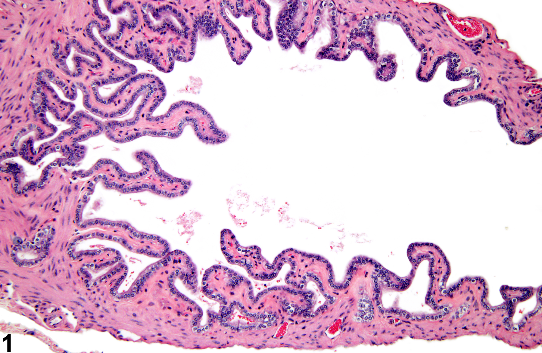
Epithelial hyperplasia in a male B6C3F1 mouse from a chronic study.
All Images

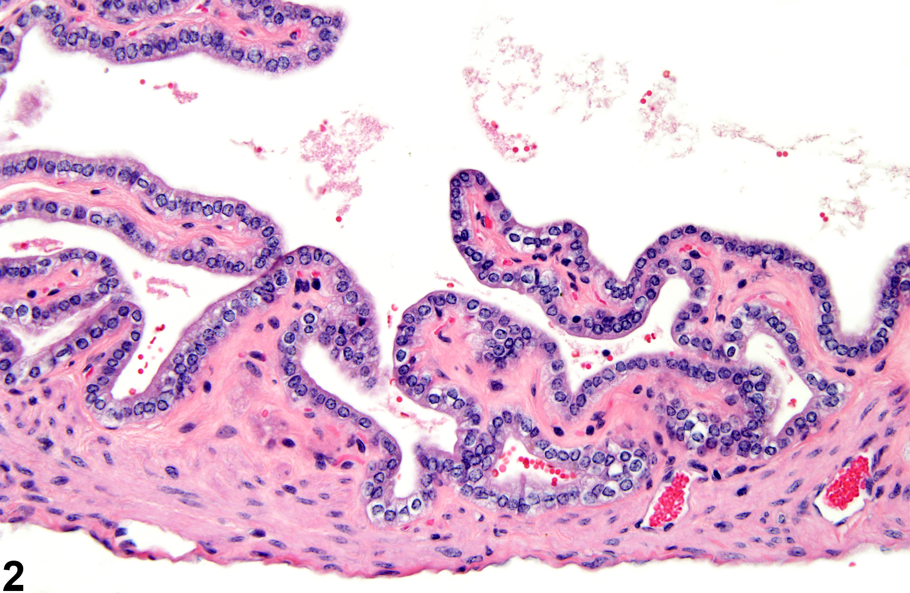
Epithelial hyperplasia in a male B6C3F1 mouse from a chronic study.

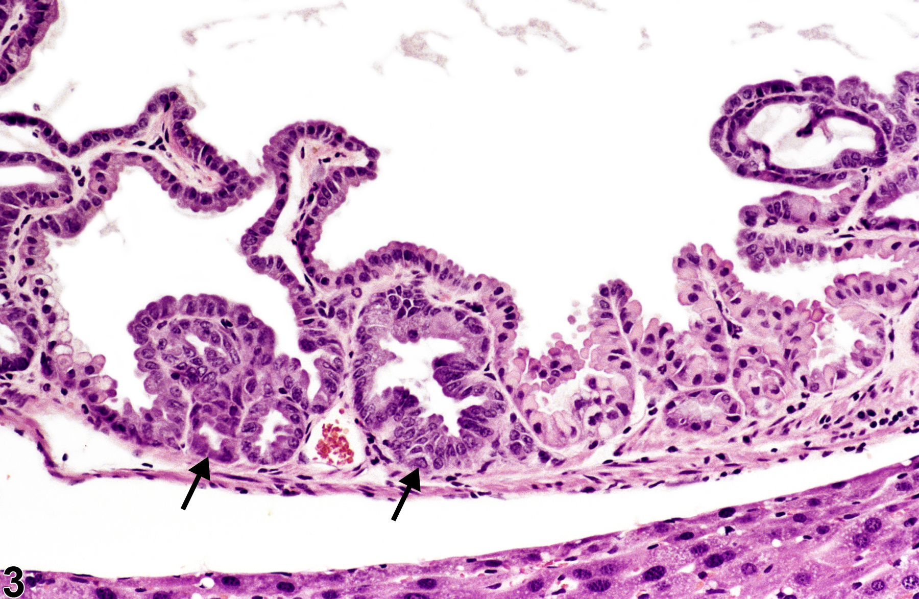
Epithelial hyperplasia in a male B6C3F1 mouse from a chronic study (higher magnification of Figure 1).

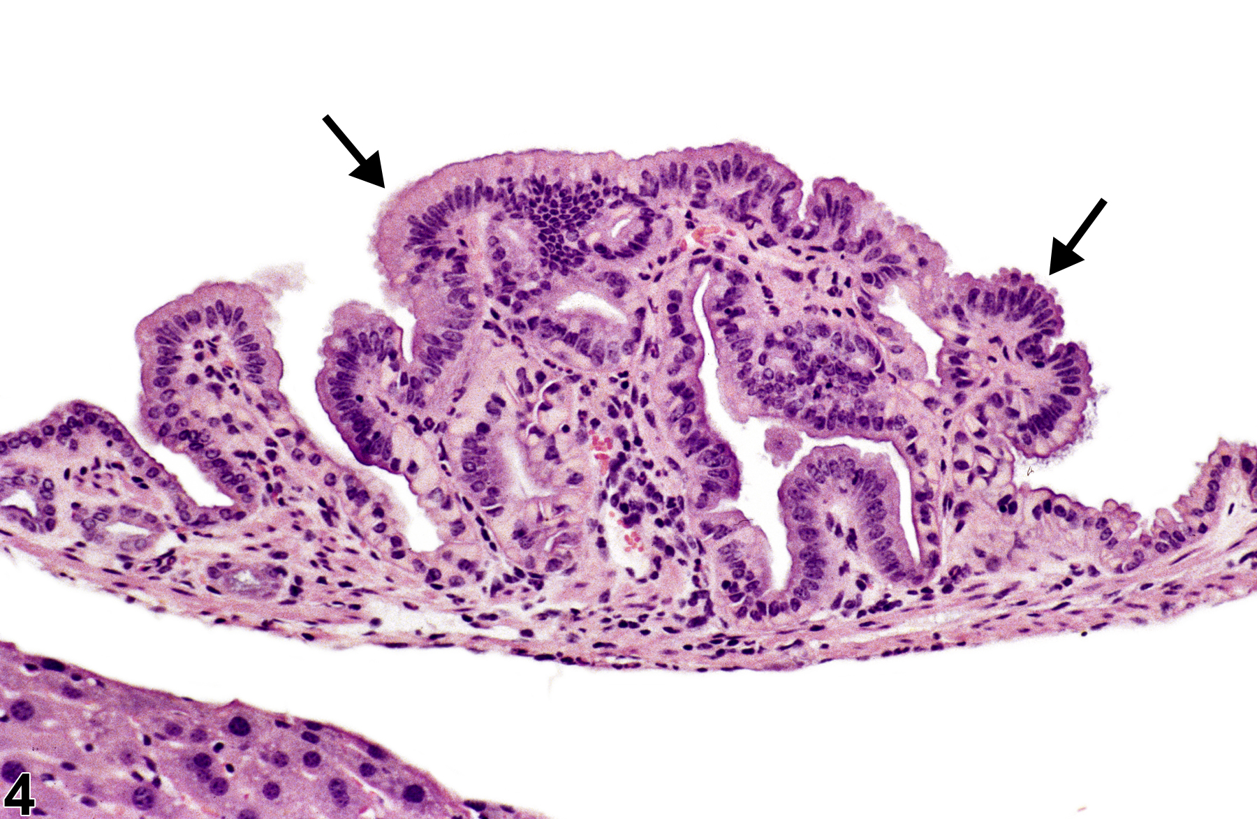
Epithelial hyperplasia-arrows indicate papillary hyperplasia in a male B6C3F1 mouse from a chronic study.

Epithelial hyperplasia-arrows indicate papillary hyperplasia in a male B6C3F1 mouse from a chronic study.