

Hepatobiliary System
Liver - Atrophy
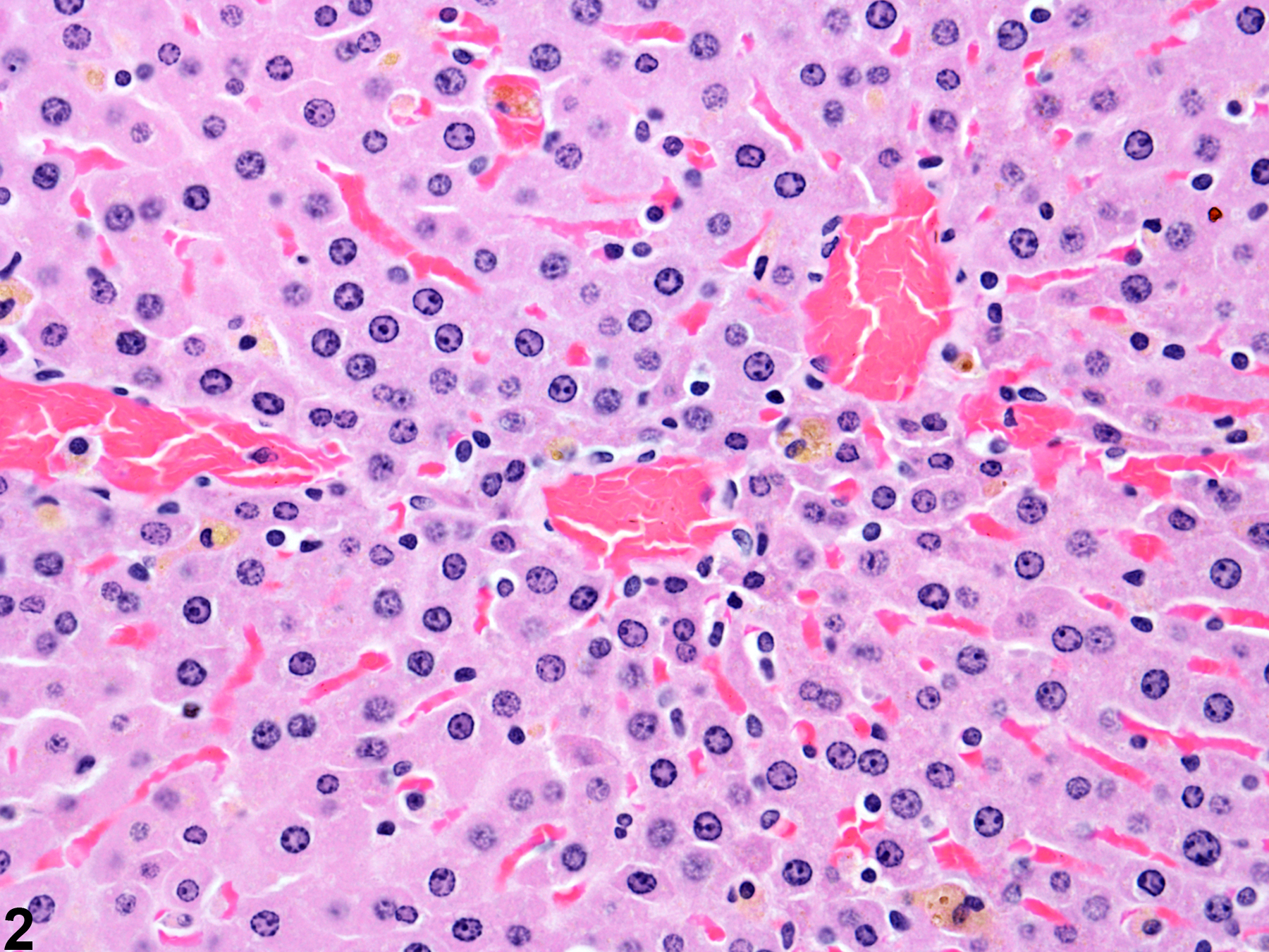
Narrative
Eustis SL, Boorman GA, Harada T, Popp JA. 1990. Liver. In: Pathology of the Fischer Rat (Boorman GA, Eustis SL, Elwell MR, Montgomery CA, MacKenzie WF, eds). Academic Press, San Diego, 71-94.
Greaves P. 2007. Histopathology of Preclinical Toxicity Studies: Interpretation and Relevance in Drug Safety Evaluation, 3rd ed. Elsevier, Amsterdam.
Abstract: http://www.sciencedirect.com/science/book/9780444527714Harada T, Enomoto A, Boorman GA, Maronpot RR. 1999. Liver and gallbladder. In: Pathology of the Mouse: Reference and Atlas (Maronpot RR, Boorman GA, Gaul BW, eds). Cache River Press, Vienna, IL, 119-183.
National Toxicology Program. 1994. NTP TOX-38. Toxicity Studies of Sodium Selenate and Sodium Selenite (CAS. No. 13410-01-0 and 10102-18-8) Administered in Drinking Water to F344/N Rats and B6C3F1 Mice. NTP, Research Triangle Park, NC.
Abstract: https://ntp.niehs.nih.gov/go/tox38absThoolen B, Maronpot RR, Harada T, Nyska A, Rousseaux C, Nolte T, Malarkey D, Kaufmann W, Kutter K, Deschl U, Nakae D, Gregson R, Winlove M, Brix A, Singl B, Belpoggi F, Ward JM. 2010. Hepatobiliary lesion nomenclature and diagnostic criteria for lesions in rats and mice (INHAND). Toxicol Pathol 38:5S-81S.
Full Text: http://tpx.sagepub.com/content/38/7_suppl/5S.full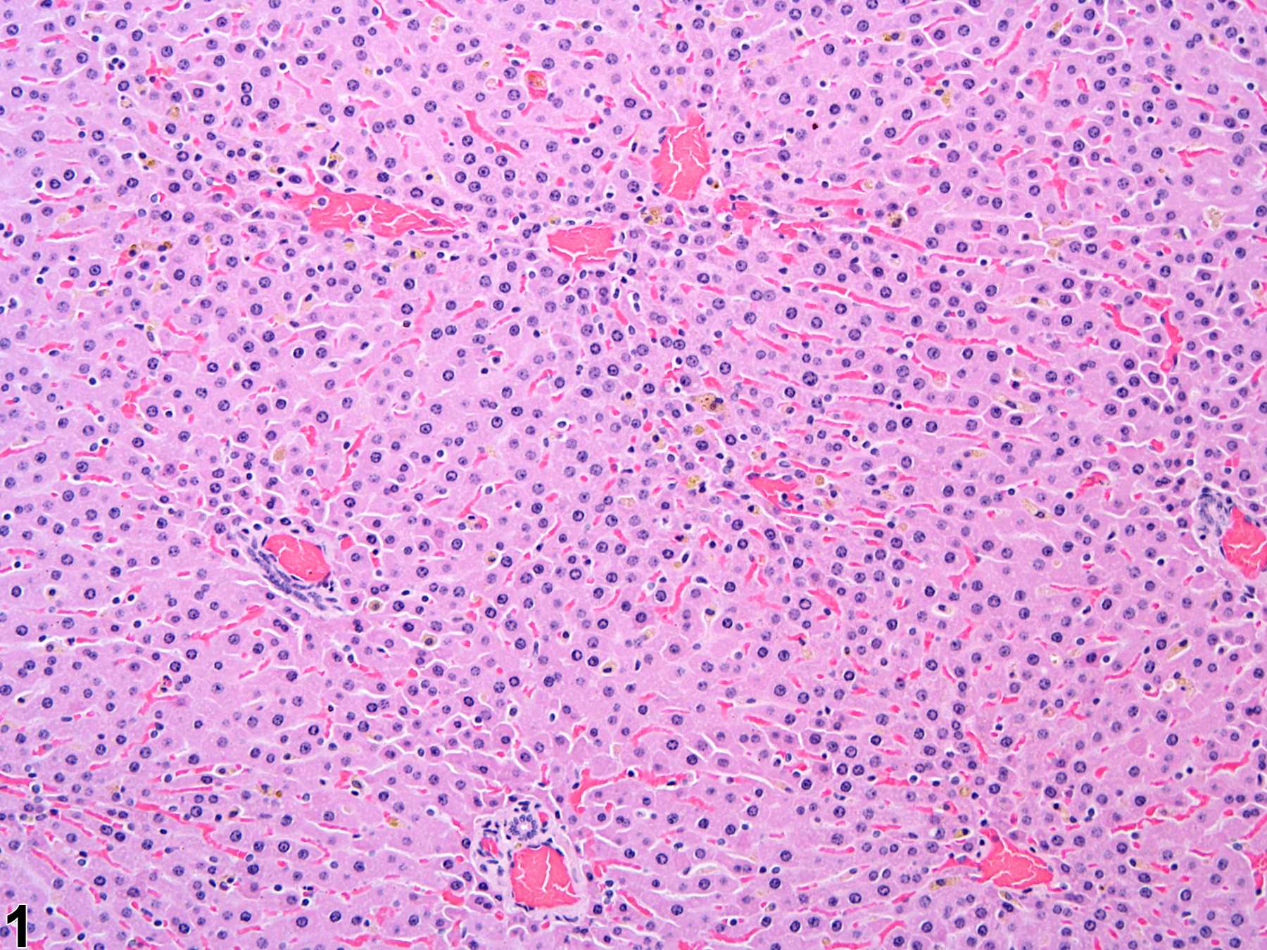
Atrophy in a female F344/N rat from a subchronic study.
All Images

Atrophy in a female F344/N rat from a subchronic study.

Atrophy in a female F344/N rat from a subchronic study.

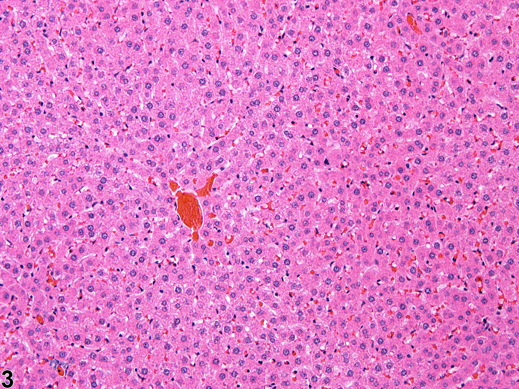
Normal liver in a control female F344/N rat from a subchronic study.

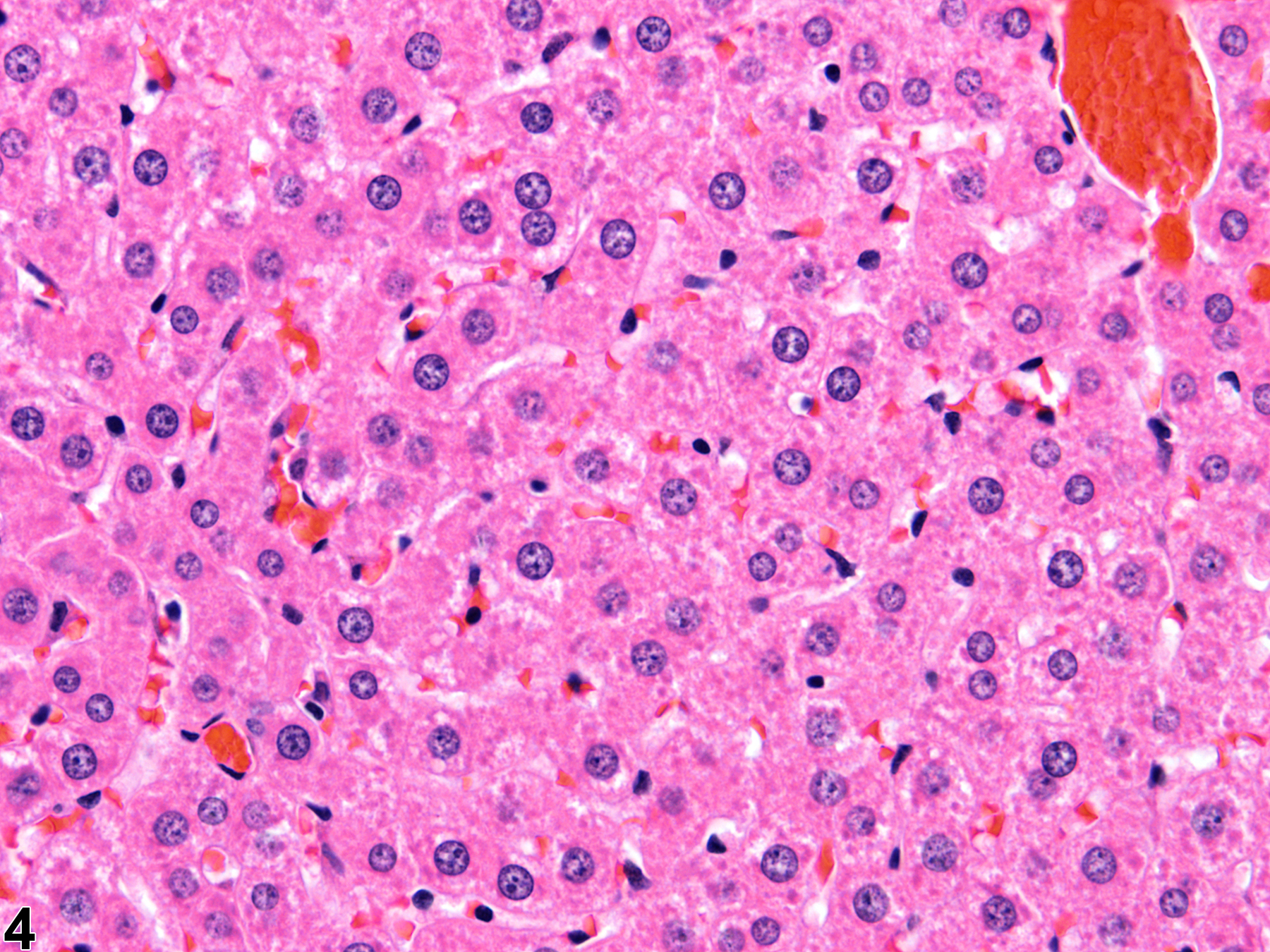
Normal liver in a control female F344/N rat from a subchronic study. Higher magnification of Figure 3.