

Hepatobiliary System
Liver, Bile Duct - Cyst
Narrative
Eustis SL, Boorman GA, Harada T, Popp JA. 1990. Liver. In: Pathology of the Fischer Rat (Boorman GA, Eustis SL, Elwell MR, Montgomery CA, MacKenzie WF, eds). Academic Press, San Diego, 71-94.
Harada T, Enomoto A, Boorman GA, Maronpot RR. 1999. Liver and gallbladder. In: Pathology of the Mouse: Reference and Atlas (Maronpot RR, Boorman GA, Gaul BW, eds). Cache River Press, Vienna, IL, 119-183.
National Toxicology Program. 2006. NTP TR-521. Toxicology and Carcinogenesis Studies of 2,3,7,8-Tetrachlorodibenzo-p-dioxin (TCDD) (CAS No. 1746-01-6) in Female Harlan Sprague-Dawley Rats (Gavage Studies). NTP, Research Triangle Park, NC.
Full Text: https://ntp.niehs.nih.gov/ntp/htdocs/lt_rpts/tr521.pdfThoolen B, Maronpot RR, Harada T, Nyska A, Rousseaux C, Nolte T, Malarkey D, Kaufmann W, Kutter K, Deschl U, Nakae D, Gregson R, Winlove M, Brix A, Singl B, Belpoggi F, Ward JM. 2010. Hepatobiliary lesion nomenclature and diagnostic criteria for lesions in rats and mice (INHAND). Toxicol Pathol 38:5S-81S.
Full Text: http://tpx.sagepub.com/content/38/7_suppl/5S.full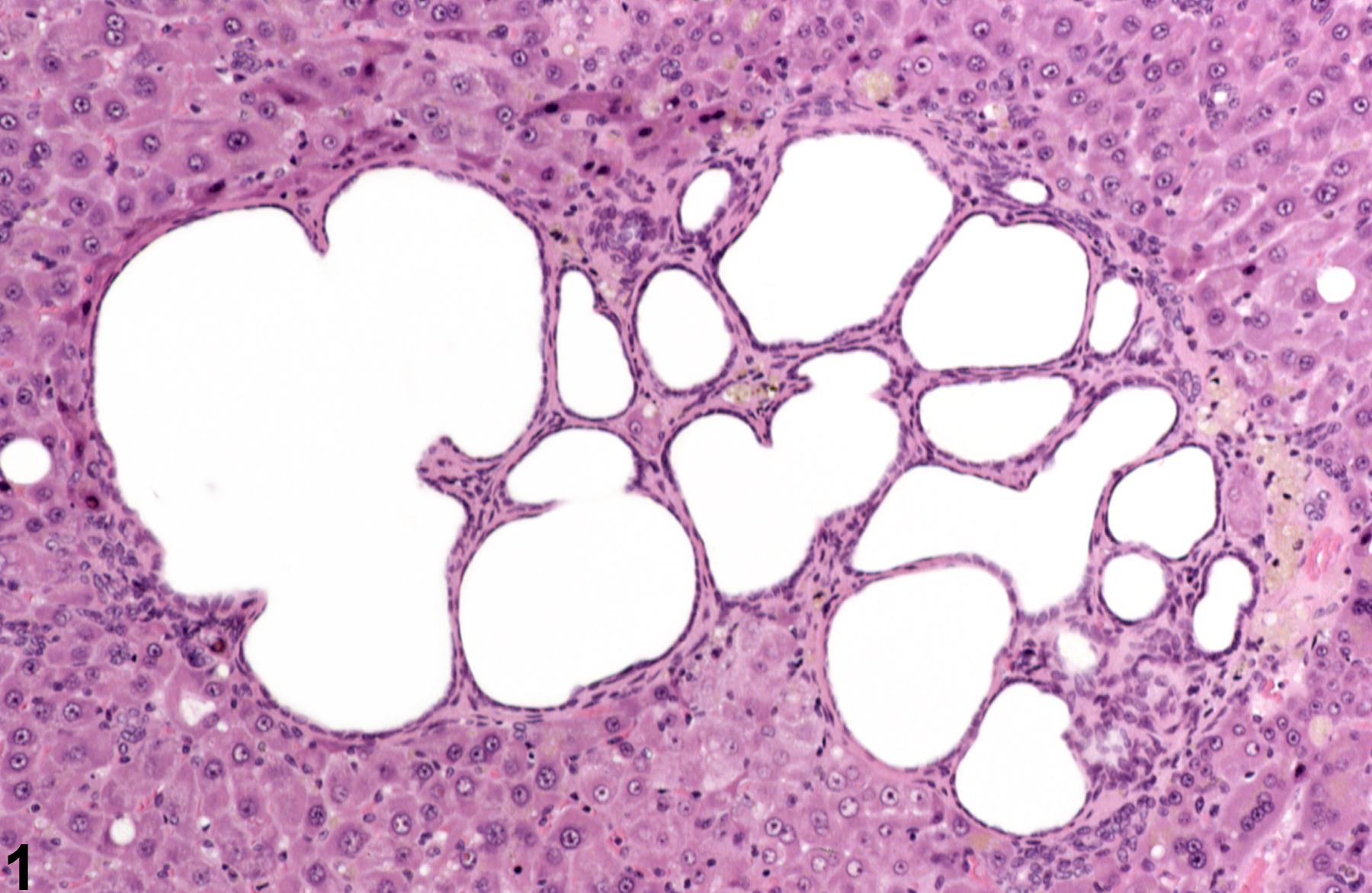
Bile duct cysts in a female Harlan Sprague-Dawley rat from a chronic study.
All Images

Bile duct cysts in a female Harlan Sprague-Dawley rat from a chronic study.

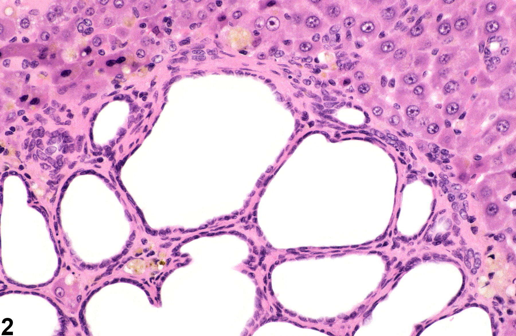
Bile duct cysts in a female Harlan Sprague-Dawley rat from a chronic study (higher magnification of Figure 1).

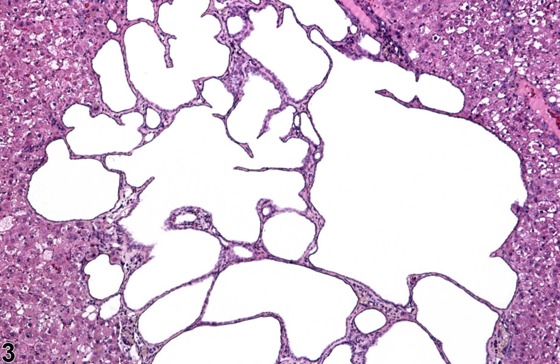
Bile duct cyst-multiloculated cyst in a female Harlan Sprague-Dawley rat from a chronic study.

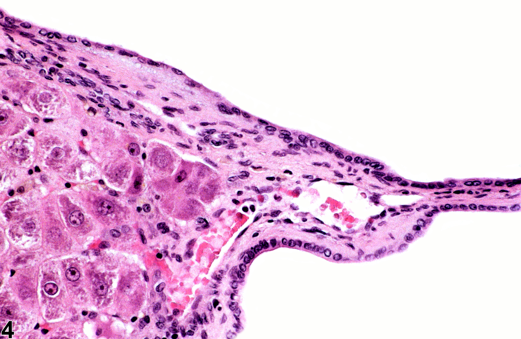
Bile duct cysts in a female Harlan Sprague-Dawley rat from a chronic study.