U.S. flag

An official website of the United States government

Dot gov

The .gov means it's official.
Federal government websites often end in .gov or .mil. Before sharing sensitive information, make sure you're on a federal government site.

Https

The site is secure.
The https:// ensures that you are connecting to the official website and that any information you provide is encrypted and transmitted securely.

Skip to Main Navigation
Skip to Page Content
Skip to Atlas Navigation

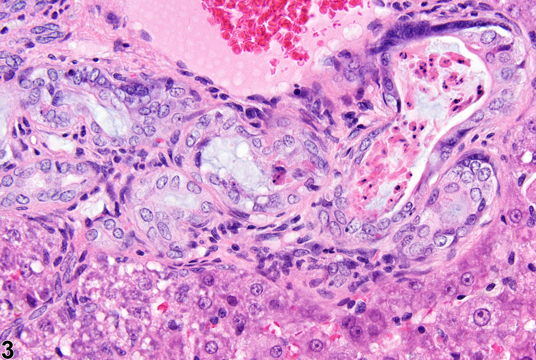
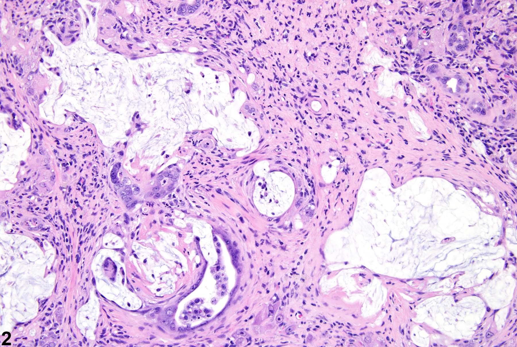
Liver - Cholangiofibrosis

Image of cholangiofibrosis in the liver from a female Harlan Sprague-Dawley rat in a chronic study
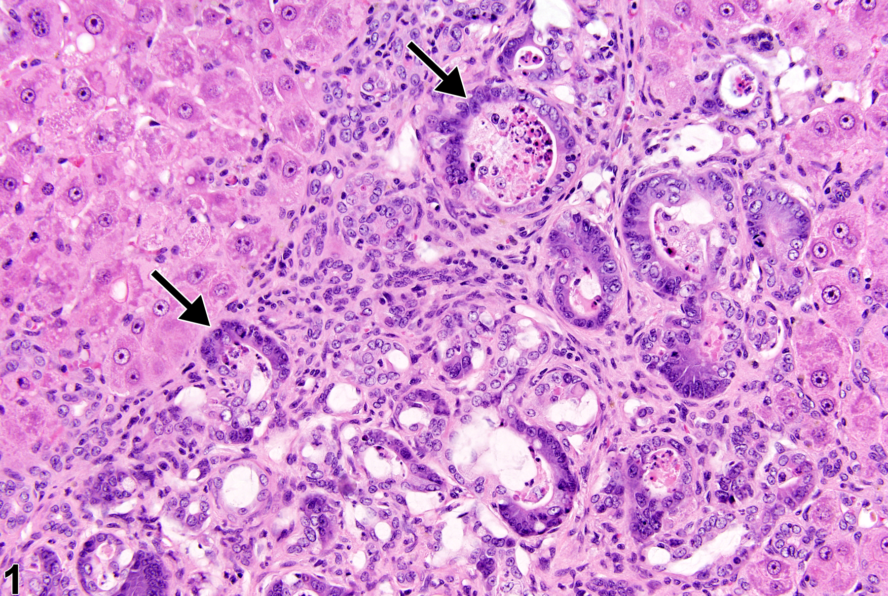
Cholangiofibrosis-arrows indicate incompletely lined proliferating bile ducts in a female Harlan Sprague-Dawley rat in a chronic study.
Figure 1 of 3
Image of cholangiofibrosis in the liver from a female Harlan Sprague-Dawley rat in a chronic study
Cholangiofibrosis in a female Harlan Sprague-Dawley rat from a chronic study.
Figure 2 of 3
Image of cholangiofibrosis in the liver from a male  F344/N rat in a subchronic study
Cholangiofibrosis in a male F344/N rat from a subchronic study.
Figure 3 of 3
next arrow

comment:

This chronic inflammatory process is initially associated with oval cell proliferation and bile duct hyperplasia with dilation of proliferating bile ducts. Bile duct contents include mucus, necrotic debris, and desquamated epithelial cells ( Figure 3image opens in a pop-up window ). Biliary structures are frequently incompletely lined (crescent-shaped bile ducts; Figure 1image opens in a pop-up window , arrows) by hyperchromatic cuboidal to columnar cells and goblet cells (intestinal metaplasia). As the lesion progresses, there is peribiliary fibrosis and a mixed inflammatory cell infiltrate ( Figure 1image opens in a pop-up window and Figure 2image opens in a pop-up window ). Large areas of hepatic lobe may be affected, with progressive resolution of lesions by development of sclerotic connective tissue, with retention of some biliary glandular acini and residual pools of mucus. Cholangiofibrosis may resemble cholangiocarcinoma but may be differentiated by the latter’s more prominent biliary epithelial changes (e.g., multiple layers or piling up of epithelial cells and pleomorphism). Epithelial cell atypia and increased mitotic figures may be present in either lesion, however, and therefore are not useful in differentiating cholangiofibrosis from cholangiocarcinoma. Some references indicate that significant bile duct dilation and intestinal metaplasia of the biliary epithelium are not prominent features of cholangiocarcinoma.

recommendation:

This inflammatory hepatic response should be documented and given a severity grade, with severity dependent on the extent of liver involvement. All the features described above are included in the diagnosis of cholangiofibrosis and should not be diagnosed separately unless they clearly represent separate processes.

related links:

Liver - Inflammation

references:

Adams ET, Auerbach S, Blackshear PE, Bradley A, Gruebbel MM, Little PB, Malarkey D, Maronpot R, McKay JS, Miller RA, Moore RR, Morrison JP, Nyska A, Ramot Y, Rao D, Suttie A, Wells MY, Willson GA, Elmore SA. 2011. Proceedings of the 2010 National Toxicology Program Satellite Symposium. Toxicol Pathol 39(1):240-266.
Abstract: https://www.ncbi.nlm.nih.gov/pubmed/21177527

Bannasch P, Zerban H. 1990 Tumours of the liver. In: Pathology of Tumours in Laboratory Animals, Vol 1: Tumours of the Rat, 2nd ed. (Turusov VS, Mohr U, eds). IARC Scientific Publication No. 99. International Agency for Research on Cancer, Lyon, France, 199-240.

Eustis SL, Boorman GA, Harada T, Popp JA. 1990. Liver. In: Pathology of the Fischer Rat (Boorman GA, Eustis SL, Elwell MR, Montgomery CA, MacKenzie WF, eds). Academic Press, San Diego, 71-94.

Hailey JR, Walker NJ, Sells DM, Brix AE, Jokinen MP, Nyska A. 2005. Classification of proliferative hepatocellular lesions in Harlan Sprague-Dawley rats chronically exposed to dioxin-like compounds. Toxicol Pathol 33:165-174.
Abstract: https://www.ncbi.nlm.nih.gov/pubmed/15805068

Kimbrough RD, Linder RE, Burse VW, Jennings RW. 1973. Adenofibrosis in the rat liver, with persistence of polychlorinated biphenyls in adipose tissue. Arch Environ Health 27:390-395.
Abstract: https://www.ncbi.nlm.nih.gov/pubmed/4356600

Sirica AE. 1992. The Role of Cell Types in Hepatocarcinogenesis. CRC Press, Boca Raton, FL.
Abstract: http://www.crcpress.com/product/isbn/9780849347467

Thoolen B, Maronpot RR, Harada T, Nyska A, Rousseaux C, Nolte T, Malarkey D, Kaufmann W, Kutter K, Deschl U, Nakae D, Gregson R, Winlove M, Brix A, Singl B, Belpoggi F, Ward JM. 2010. Hepatobiliary lesion nomenclature and diagnostic criteria for lesions in rats and mice (INHAND). Toxicol Pathol 38:5S-81S.
Full Text: http://tpx.sagepub.com/content/38/7_suppl/5S.full