

Hepatobiliary System
Liver - Fatty Change
Narrative
Fatty change is frequently accompanied by other cytoplasmic alterations, such as glycogen accumulation or depletion, and it can be difficult to distinguish the different types of alterations in cytoplasmic morphology. In those situations, a diagnosis of hepatocyte cytoplasmic vacuolization is appropriate, along with a severity grade, but the morphology of the cytoplasmic changes in the hepatocytes should be thoroughly described in the pathology narrative. The pathology narrative should also include the pathologist’s opinions regarding the contents of the cytoplasmic vacuoles.
Cattley RC, Popp JA. 2002. Liver. In: Handbook of Toxicologic Pathology (Haschek WM, Rousseaux CG, Wallig MA, eds). Academic Press, San Diego, 2:187-225.
Eustis SL, Boorman GA, Harada T, Popp JA. 1990. Liver. In: Pathology of the Fischer Rat (Boorman GA, Eustis SL, Elwell MR, Montgomery CA, MacKenzie WF, eds). Academic Press, San Diego, 71-94.
Evans JG, Lake BG. 1998. The digestive system II. Hepatobiliary system. In: Target Organ Pathology (Turton J, Hooson J, eds). Taylor and Francis, London, 61-98.
Greaves P. 2007. Histopathology of Preclinical Toxicity Studies: Interpretation and Relevance in Drug Safety Evaluation, 3rd ed. Elsevier, Amsterdam.
Abstract: http://www.sciencedirect.com/science/book/9780444527714Harada T, Enomoto A, Boorman GA, Maronpot RR. 1999. Liver and gallbladder. In: Pathology of the Mouse: Reference and Atlas (Maronpot RR, Boorman GA, Gaul BW, eds). Cache River Press, Vienna, IL, 119-183.
Hardisty JF, Brix AE. 2005. Comparative hepatic toxicity: Prechronic/chronic liver toxicity in rodents. Toxicol Pathol 33:35-40.
Full Text: http://tpx.sagepub.com/content/33/1/35.full.pdfHaschek WM, Rousseaux CG, Wallig MA. 2010. Fundamentals of Toxicologic Pathology, 2nd ed. Academic Press, San Diego, 197-235.
Thoolen B, Maronpot RR, Harada T, Nyska A, Rousseaux C, Nolte T, Malarkey D, Kaufmann W, Kutter K, Deschl U, Nakae D, Gregson R, Winlove M, Brix A, Singl B, Belpoggi F, Ward JM. 2010. Hepatobiliary lesion nomenclature and diagnostic criteria for lesions in rats and mice (INHAND). Toxicol Pathol 38:5S-81S.
Full Text: http://tpx.sagepub.com/content/38/7_suppl/5S.full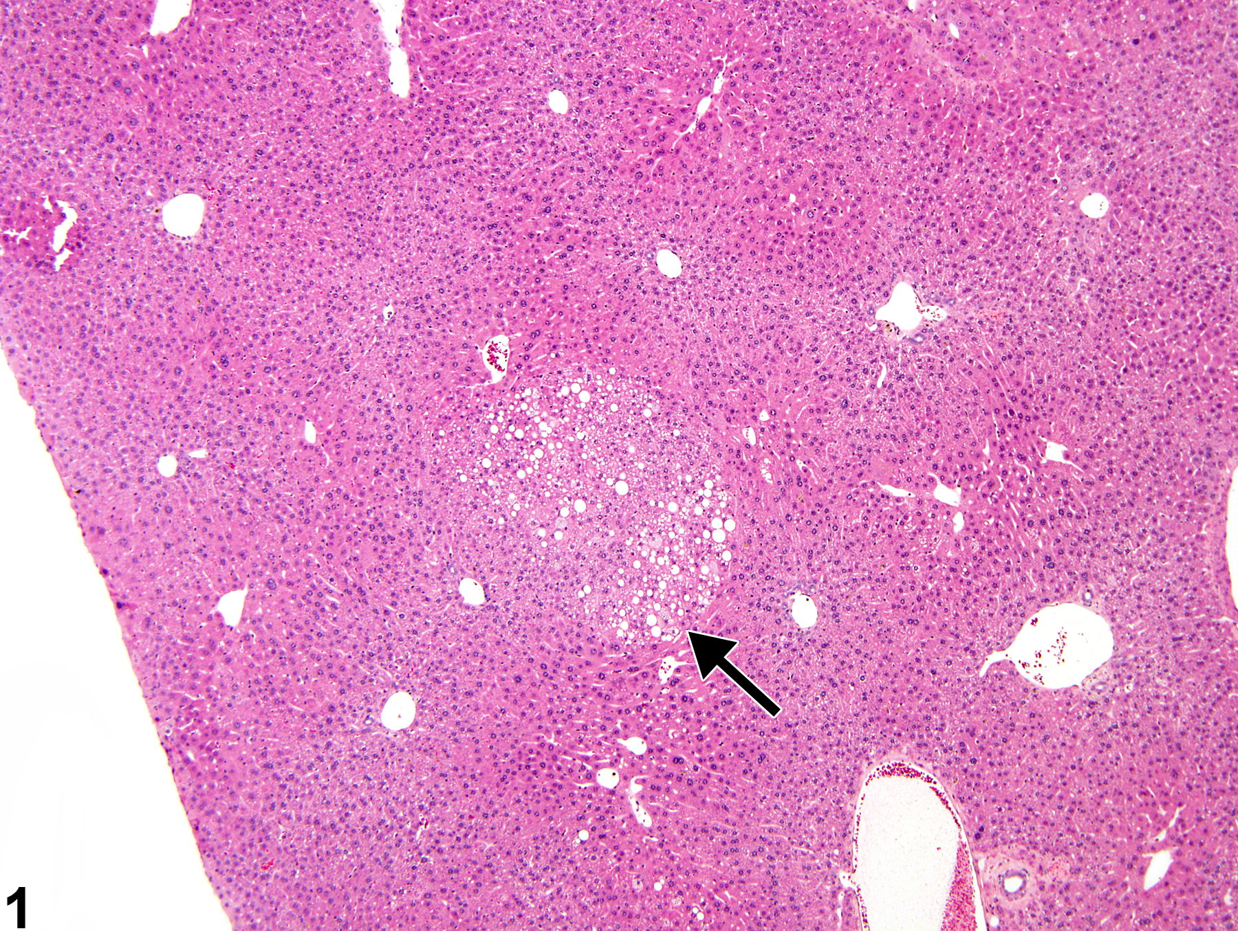
Fatty change-focal fatty change (arrow) in a B6C3F1 male mouse from a chronic study.
All Images

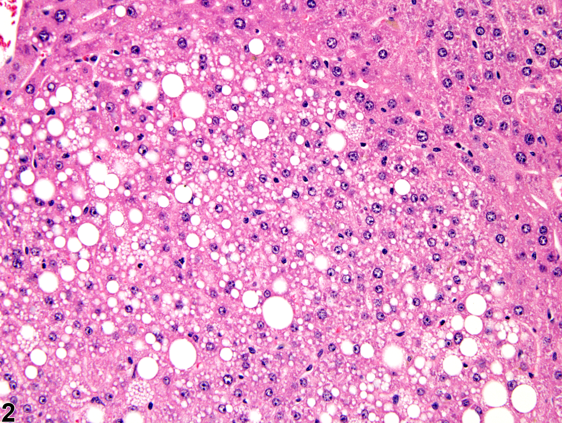
Fatty change-focal fatty change (arrow) in a B6C3F1 male mouse from a chronic study.

Fatty change-focal fatty change in a B6C3F1 male mouse from a chronic study (higher magnification of Figure 1).

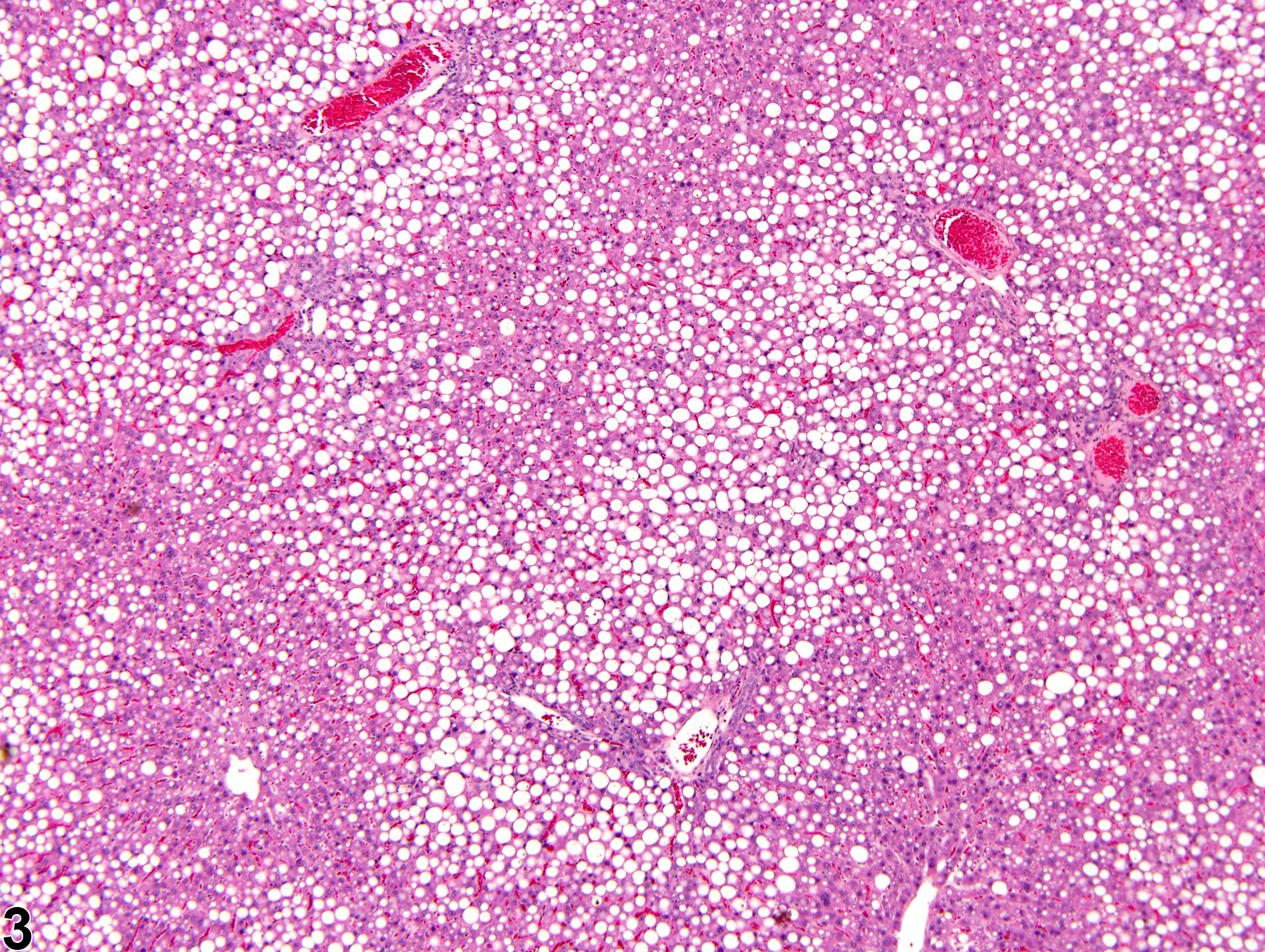
Fatty change-macrovesicular fatty change in a male F344/N rat from a chronic study.

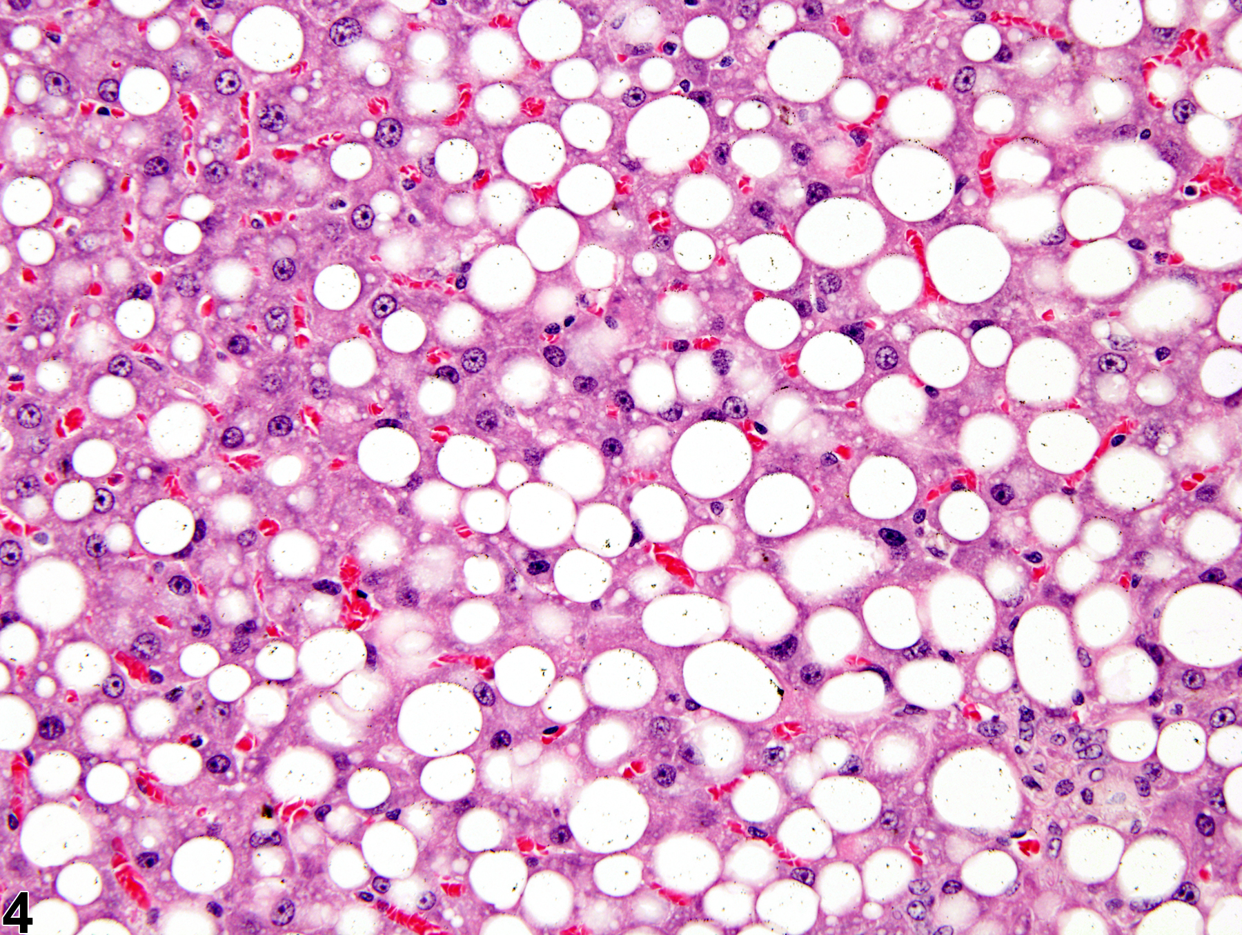
Fatty change-macrovesicular fatty change in a male F344/N rat from a chronic study (higher magnification of Figure 3).

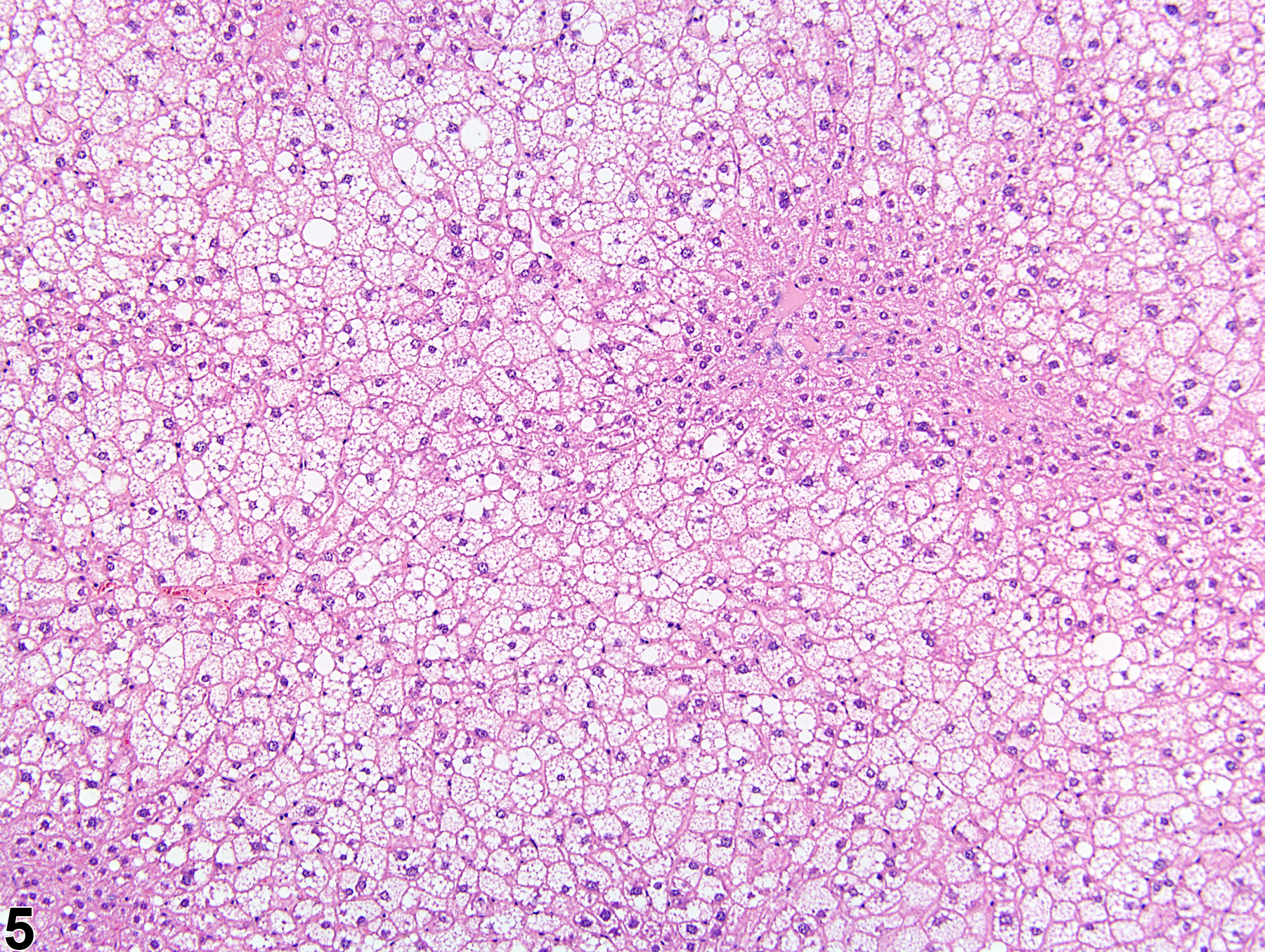
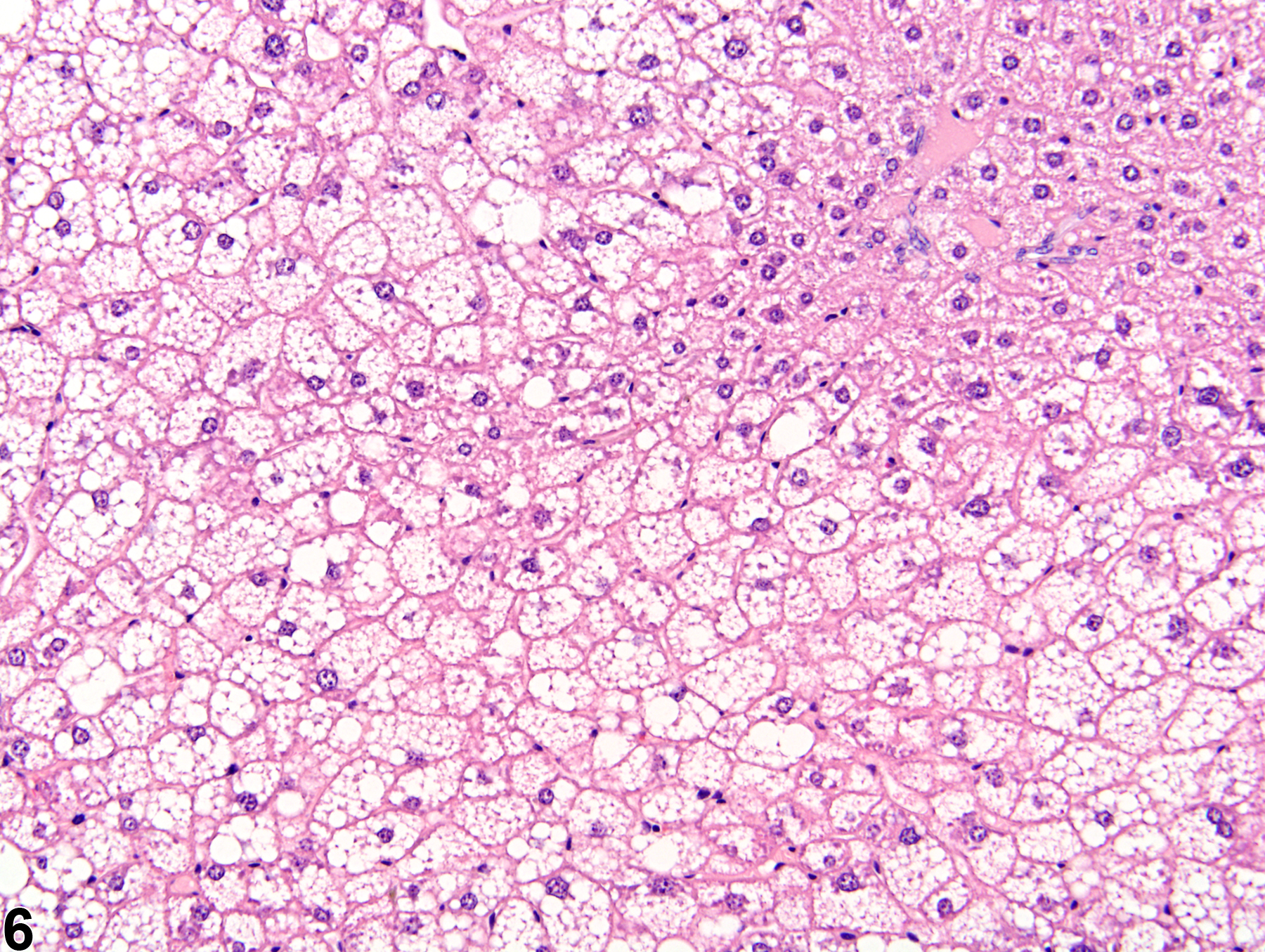
Fatty change-microvesicular fatty change in a male P53+/- (C57Bl/6) mouse from a chronic study.

Fatty change-microvesicular fatty change in a male P53+/- (C57Bl/6) mouse from a chronic study (higher magnification of Figure 5).

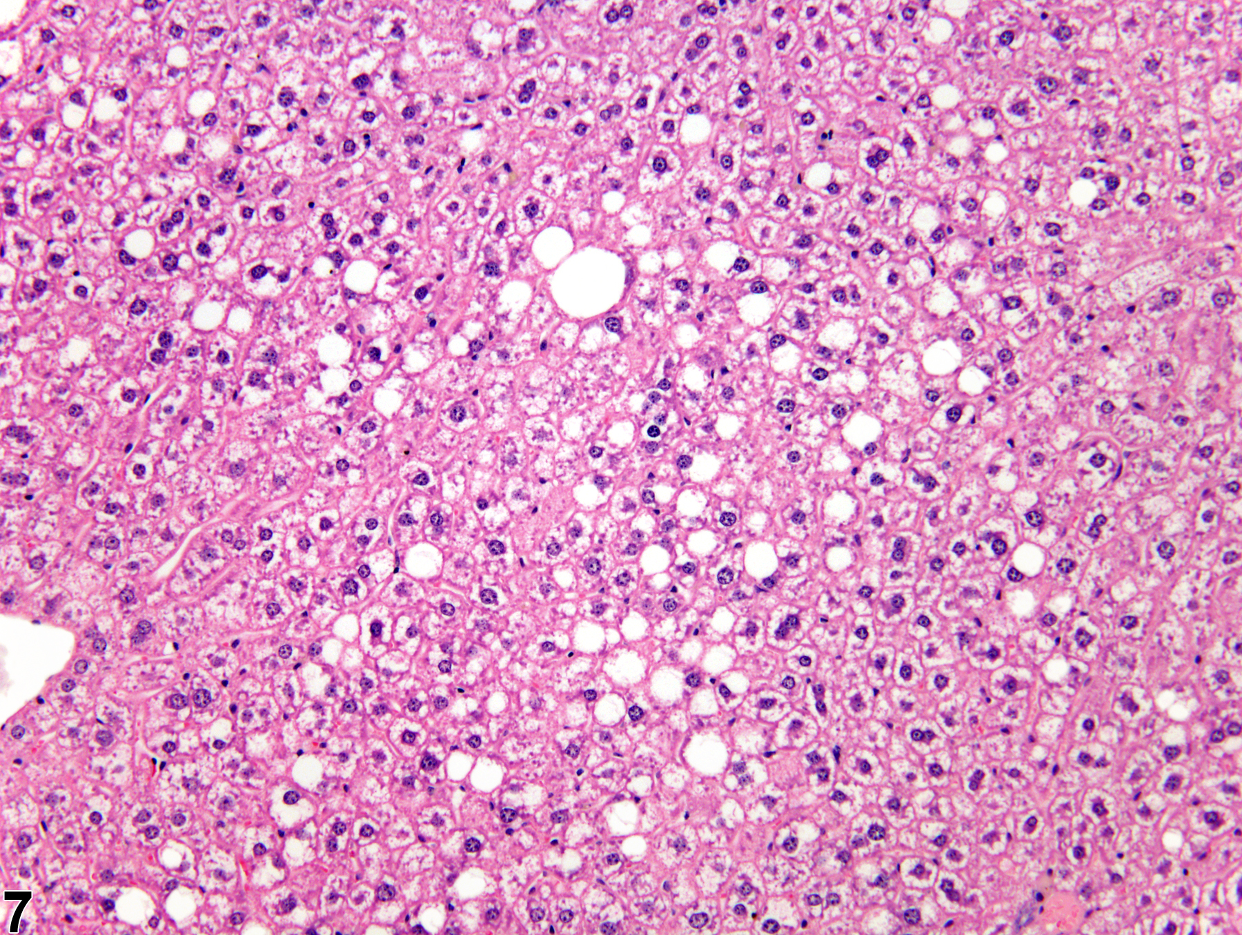
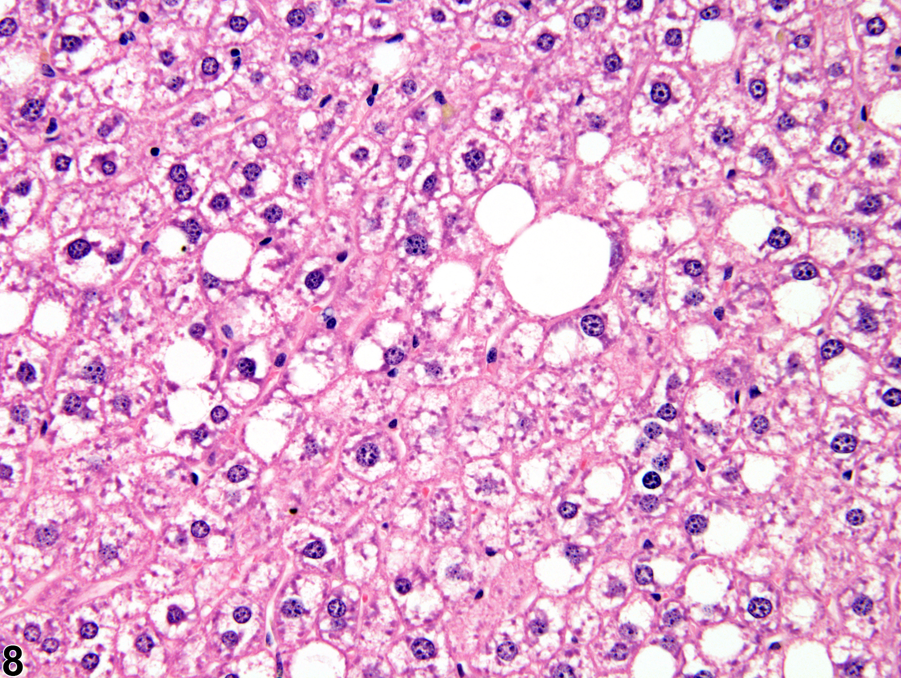
Fatty change-hepatocyte cytoplasmic vacuolation consistent with glycogen and macrovesicular fatty change in a male B6C3F1 mouse from a chronic study.

Fatty change-hepatocyte cytoplasmic vacuolation consistent with glycogen and macrovesicular fatty change in a male B6C3F1 mouse from a chronic study (higher magnification of Figure 7).