Liver, Hepatocyte - Apoptosis

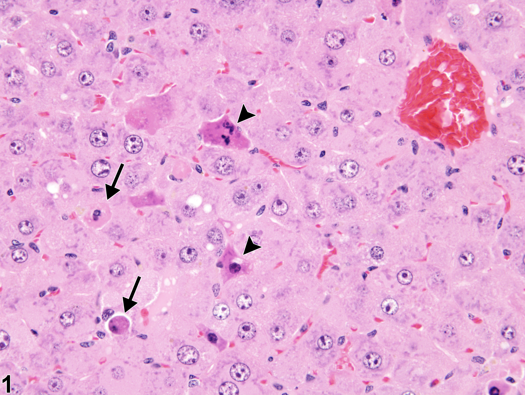
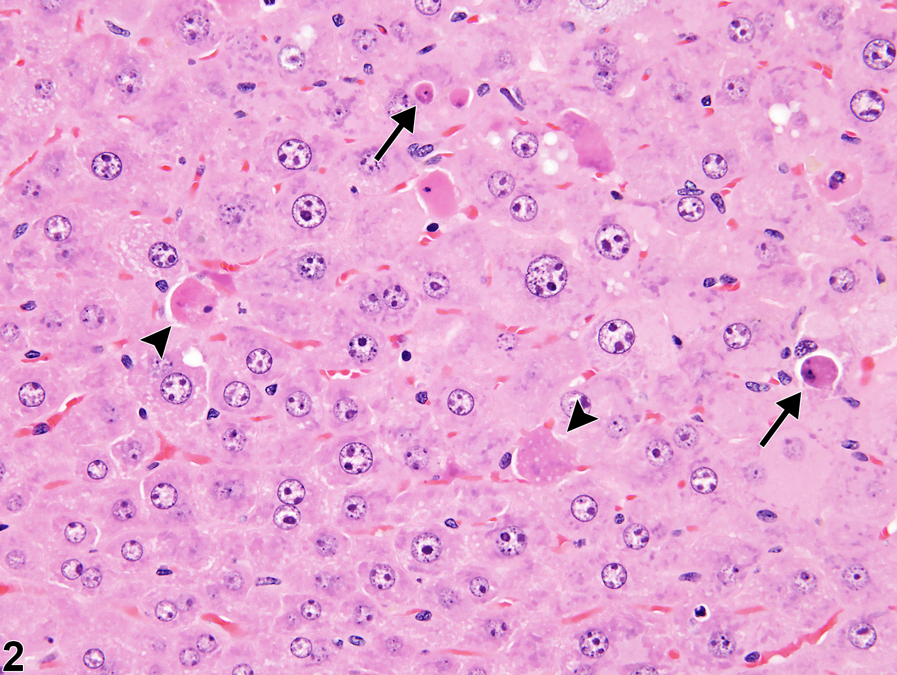
Liver, Hepatocyte - Apoptosis in a male B6C3F1/N mouse from a subchronic study. There are slightly shrunken, irregularly shaped cells consistent with early-stage apoptosis (arrowheads) and smaller, rounded cells consistent with late-stage apoptosis (arrows); both have hypereosinophilic cytoplasm and small, fragmented nuclei.
Figure 1 of 2
comment:
It is necessary to recognize that single-cell death of hepatocytes usually occurs by apoptosis. Cell death in the liver manifests as a spectrum of morphological patterns that can occur alone or in combinations. However, cell death has two primary manifestations: necrosis and apoptosis. Apoptosis represents a form of energy-dependent cell death (programmed cell death) that typically occurs as death of individual hepatocytes. In contrast to necrosis, apoptosis is a regulated and genetically controlled process. Apoptosis of individual hepatocytes may occur spontaneously as hepatocytes age and are replaced, or following injury such as exposure to hepatotoxic xenobiotics or low oxygen tension in the circulating blood. Usually no inflammation is associated with apoptosis. Affected cells have condensed, deeply eosinophilic cytoplasm in contrast to adjacent normal hepatocytes and either are devoid of chromatin or contain pyknotic or fragmented nuclear material. When affected, apoptotic hepatocytes may be small, rounded, and surrounded by a clear halo. This change is morphologically compatible with an advance stage of apoptosis, and the small cell fragments are referred to as apoptotic bodies ( Figure 1


recommendation:
Apoptosis should be diagnosed when present and given a severity grade based on the number of apoptotic cells. Although difficult to distinguish from necrosis, apoptosis can also be found at the peripheral edges of some regions of confluent coagulation necrosis. In this situation the predominant change of necrosis should be diagnosed. If necrosis occurs at high doses of a xenobiotic but apoptosis is seen at low doses with the same xenobiotic and within the same study, then the most abundant form of cell death should be diagnosed for each dose group and animal within a group and a proposed interpretation given in the pathology narrative.references:
Web page last updated on: November 29, 2016