U.S. flag

An official website of the United States government

Dot gov

The .gov means it's official.
Federal government websites often end in .gov or .mil. Before sharing sensitive information, make sure you're on a federal government site.

Https

The site is secure.
The https:// ensures that you are connecting to the official website and that any information you provide is encrypted and transmitted securely.

Skip to Main Navigation
Skip to Page Content
Skip to Atlas Navigation

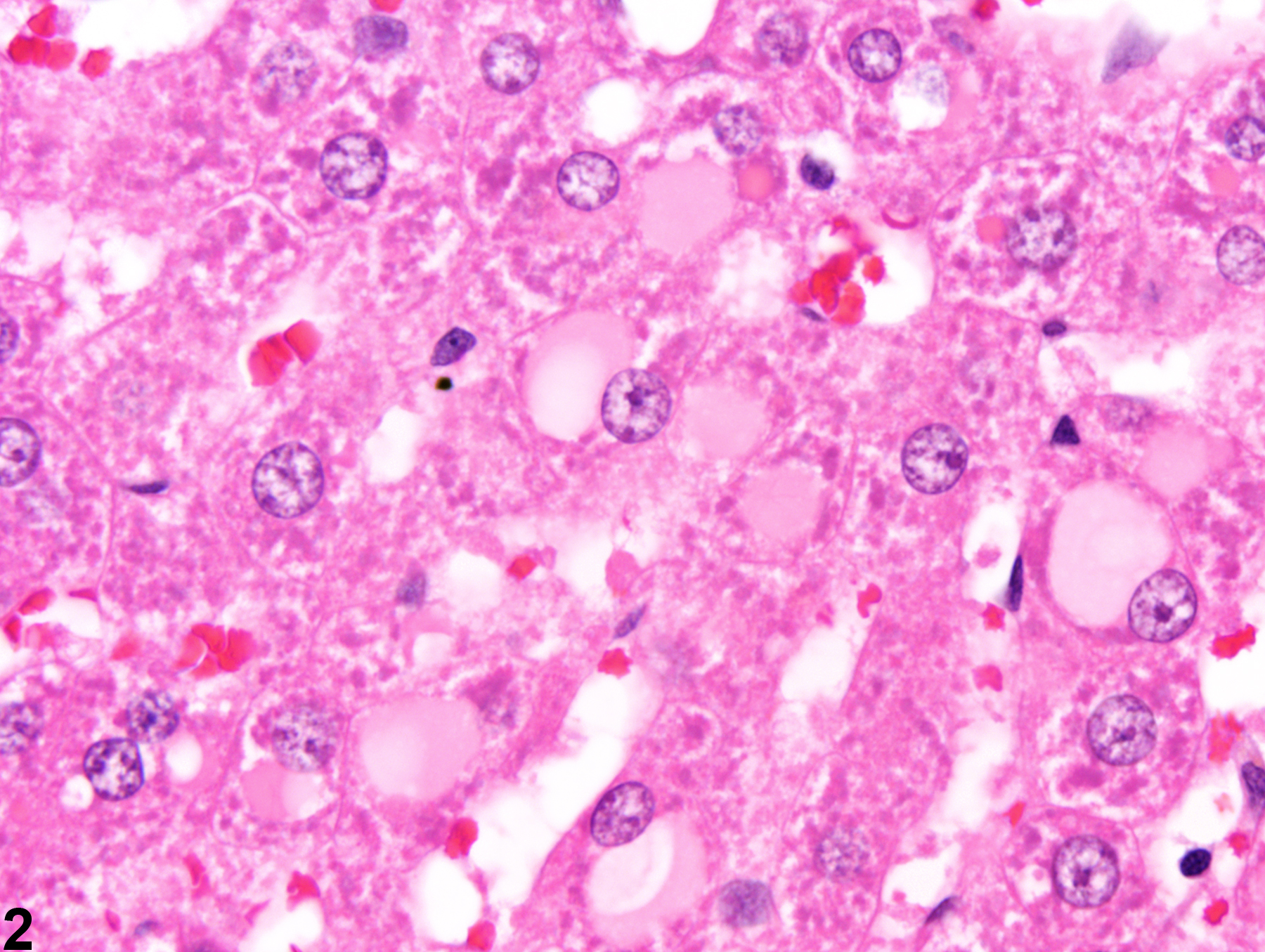
Liver, Hepatocyte - Cytoplasmic Inclusions

Image of cytoplasmic inclusions in the liver from a male  F344/N rat in a subchronic study
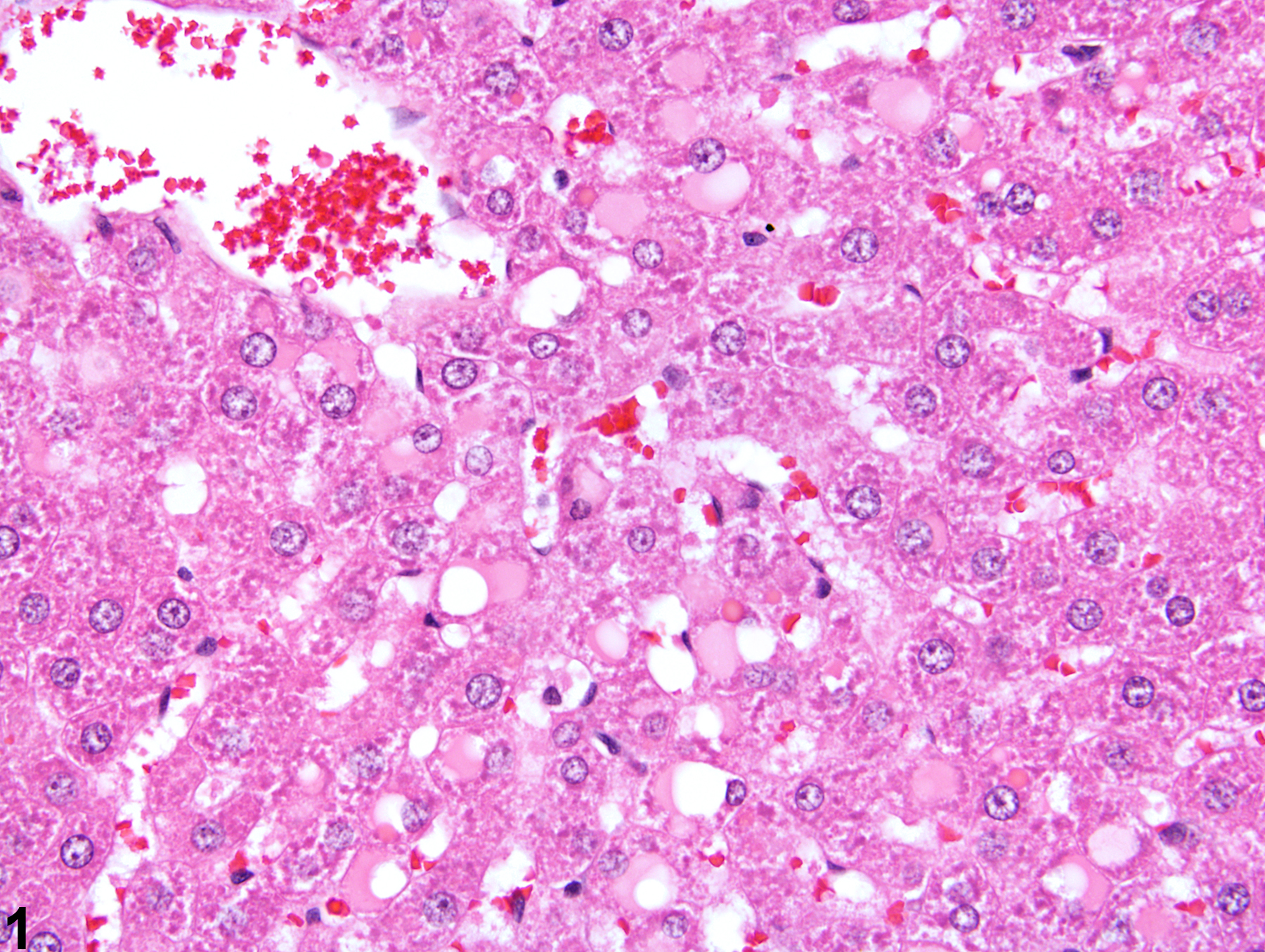
Cytoplasmic inclusions in a male F344/N rat from a subchronic study.
Figure 1 of 2
Image of cytoplasmic inclusions in the liver from a male  F344/N rat in a subchronic study
Cytoplasmic inclusions (Figure 2, arrows) in a male F344/N rat from a subchronic study.
Figure 2 of 2
next arrow

comment:

Eosinophilic or cytoplasmic inclusions or material ( Figure 2image opens in a pop-up window , arrows) can be found in control as well as treated rodents. These hyalinized inclusions are also seen in hepatocellular neoplasms. The examples in Figure 1image opens in a pop-up window and Figure 2image opens in a pop-up window represent a postmortem, time-dependent artifact especially common in male rats that are not completely exsanguinated at necropsy. The cytoplasmic vacuoles contain plasma that has entered the hepatocyte cytoplasm (plasma influx) of affected hepatocytes (Li et al. 2003). Similar but more discrete eosinophilic hyalinized inclusions, which may represent protein present in lysosomes, can be seen in control and treated rodents and are also seen in hepatocellular neoplasms.

recommendation:

Hyaline intracytoplasmic inclusions are relatively uncommon and thus should be documented and given a severity grade that is based on the frequency of affected hepatocytes. If there is reason to believe that they are a time-dependent artifact associated with incomplete exsanguination at necropsy, this should be discussed in the pathology narrative. Cytoplasmic inclusions need not be documented when present in neoplastic hepatocytes.

references:

Eustis SL, Boorman GA, Harada T, Popp JA. 1990. Liver. In: Pathology of the Fischer Rat (Boorman GA, Eustis SL, Elwell MR, Montgomery CA, MacKenzie WF, eds). Academic Press, San Diego, 71-94.

Greaves P. 2007. Histopathology of Preclinical Toxicity Studies: Interpretation and Relevance in Drug Safety Evaluation, 3rd ed. Elsevier, Amsterdam.
Abstract: http://www.sciencedirect.com/science/book/9780444527714

Harada T, Enomoto A, Boorman GA, Maronpot RR. 1999. Liver and gallbladder. In: Pathology of the Mouse: Reference and Atlas (Maronpot RR, Boorman GA, Gaul BW, eds). Cache River Press, Vienna, IL, 119-183.
Abstract: http://www.cacheriverpress.com/books/pathmouse.htm

Li X, Elwell MR, Ryan AM, Ochoa R. 2003. Morphogenesis of postmortem hepatocyte vacuolation and liver weight increases in Sprague-Dawley rats. Toxicol Pathol 31:682-688.
Abstract: https://www.ncbi.nlm.nih.gov/pubmed/14585737

Thoolen B, Maronpot RR, Harada T, Nyska A, Rousseaux C, Nolte T, Malarkey D, Kaufmann W, Kutter K, Deschl U, Nakae D, Gregson R, Winlove M, Brix A, Singl B, Belpoggi F, Ward JM. 2010. Hepatobiliary lesion nomenclature and diagnostic criteria for lesions in rats and mice (INHAND). Toxicol Pathol 38:5S-81S.
Full Text: http://tpx.sagepub.com/content/38/7_suppl/5S.full