

Hepatobiliary System
Liver - Intrahepatocellular Erythrocytes
Narrative
Barni S, Bernocchi G. 1991. Internalization of erythrocytes into liver parenchymal cells in naturally hibernating frogs (Rana esculenta L.). J Exp Zool 258:143-150.
Abstract: https://www.ncbi.nlm.nih.gov/pubmed/2022945Harada T, Enomoto A, Boorman GA, Maronpot RR. 1999. Liver and gallbladder. In: Pathology of the Mouse: Reference and Atlas (Maronpot RR, Boorman GA, Gaul BW, eds). Cache River Press, Vienna, IL, 119-183.
Lee KP. 1983. Peliosis hepatis-like lesion in aging rats. Vet Pathol 20:410-423.
Abstract: https://www.ncbi.nlm.nih.gov/pubmed/6623845Thoolen B, Maronpot RR, Harada T, Nyska A, Rousseaux C, Nolte T, Malarkey D, Kaufmann W, Kutter K, Deschl U, Nakae D, Gregson R, Winlove M, Brix A, Singl B, Belpoggi F, Ward JM. 2010. Hepatobiliary lesion nomenclature and diagnostic criteria for lesions in rats and mice (INHAND). Toxicol Pathol 38:5S-81S.
Full Text: http://tpx.sagepub.com/content/38/7_suppl/5S.full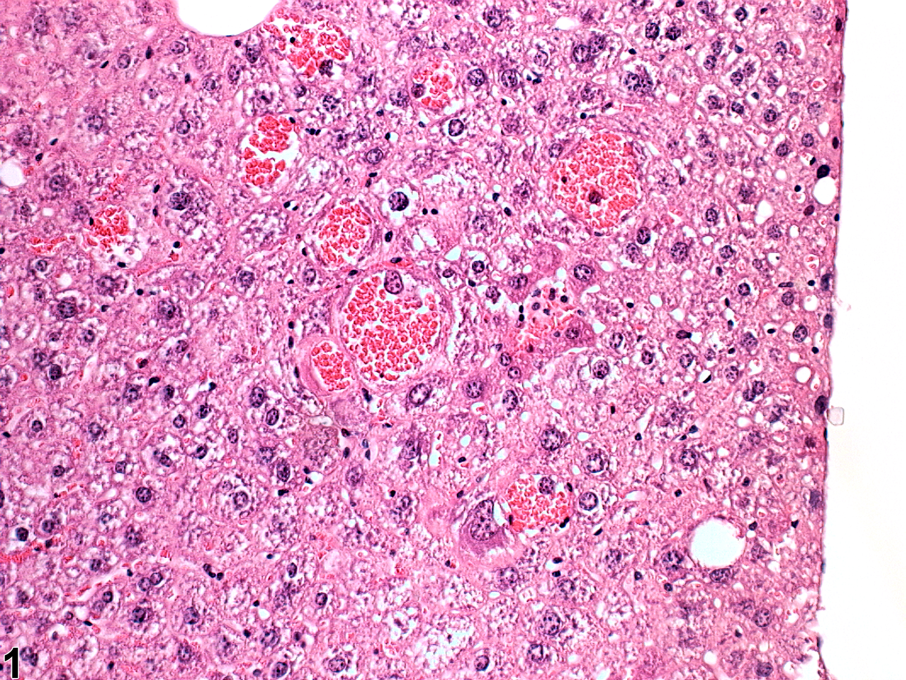
Intrahepatocellular erythrocytes in a B6C3F1 mouse from a 2-year study.
All Images

Intrahepatocellular erythrocytes in a B6C3F1 mouse from a 2-year study.
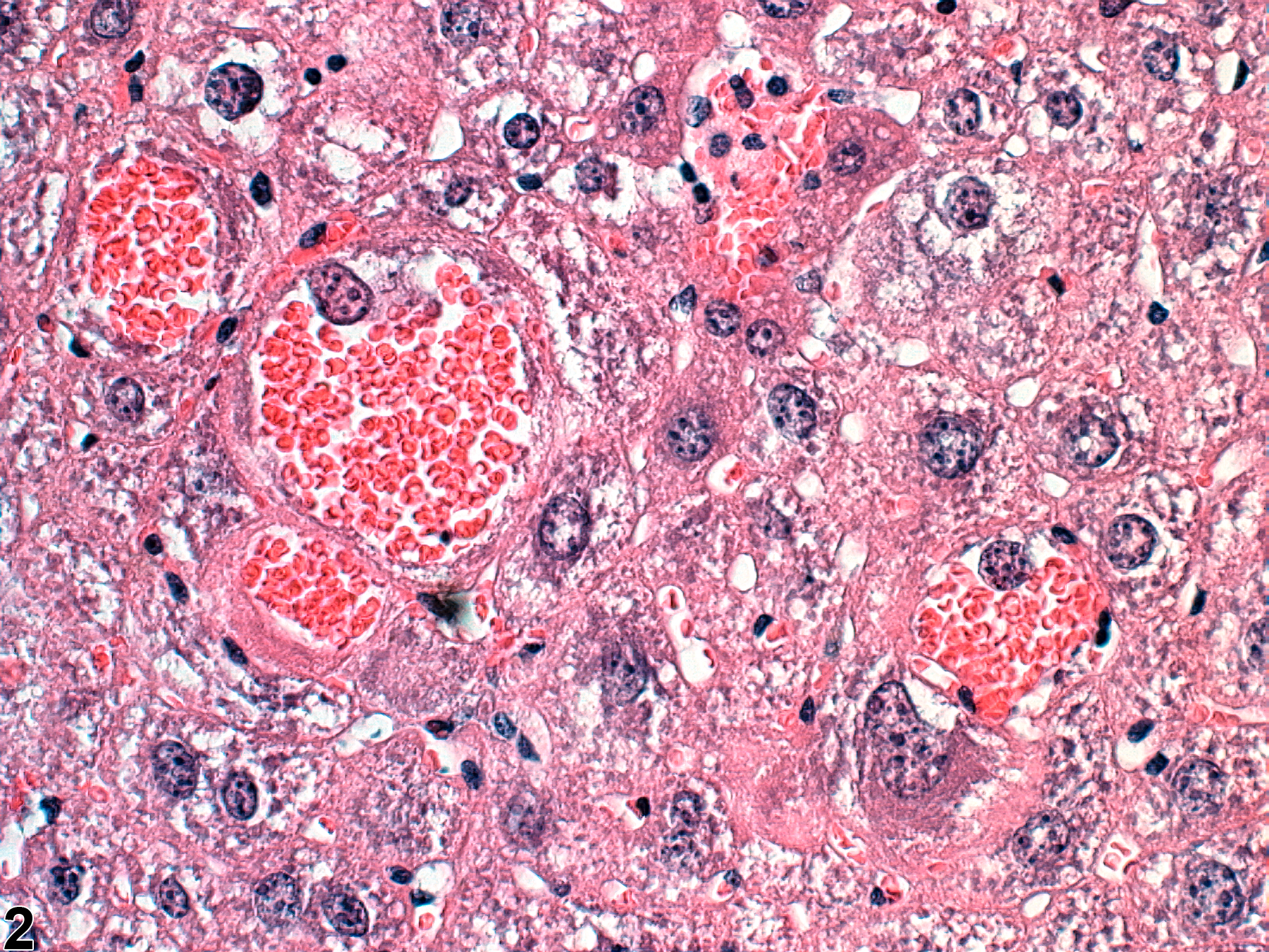
Intrahepatocellular erythrocytes in a B6C3F1 mouse from a 2-year study (higher magnification of Figure 1).