U.S. flag

An official website of the United States government

Dot gov

The .gov means it's official.
Federal government websites often end in .gov or .mil. Before sharing sensitive information, make sure you're on a federal government site.

Https

The site is secure.
The https:// ensures that you are connecting to the official website and that any information you provide is encrypted and transmitted securely.

Skip to Main Navigation
Skip to Page Content
Skip to Atlas Navigation

Liver, Stellate Cell - Hyperplasia

Image of hyperplasia in the liver from a female B6C3F1 mouse in a chronic study
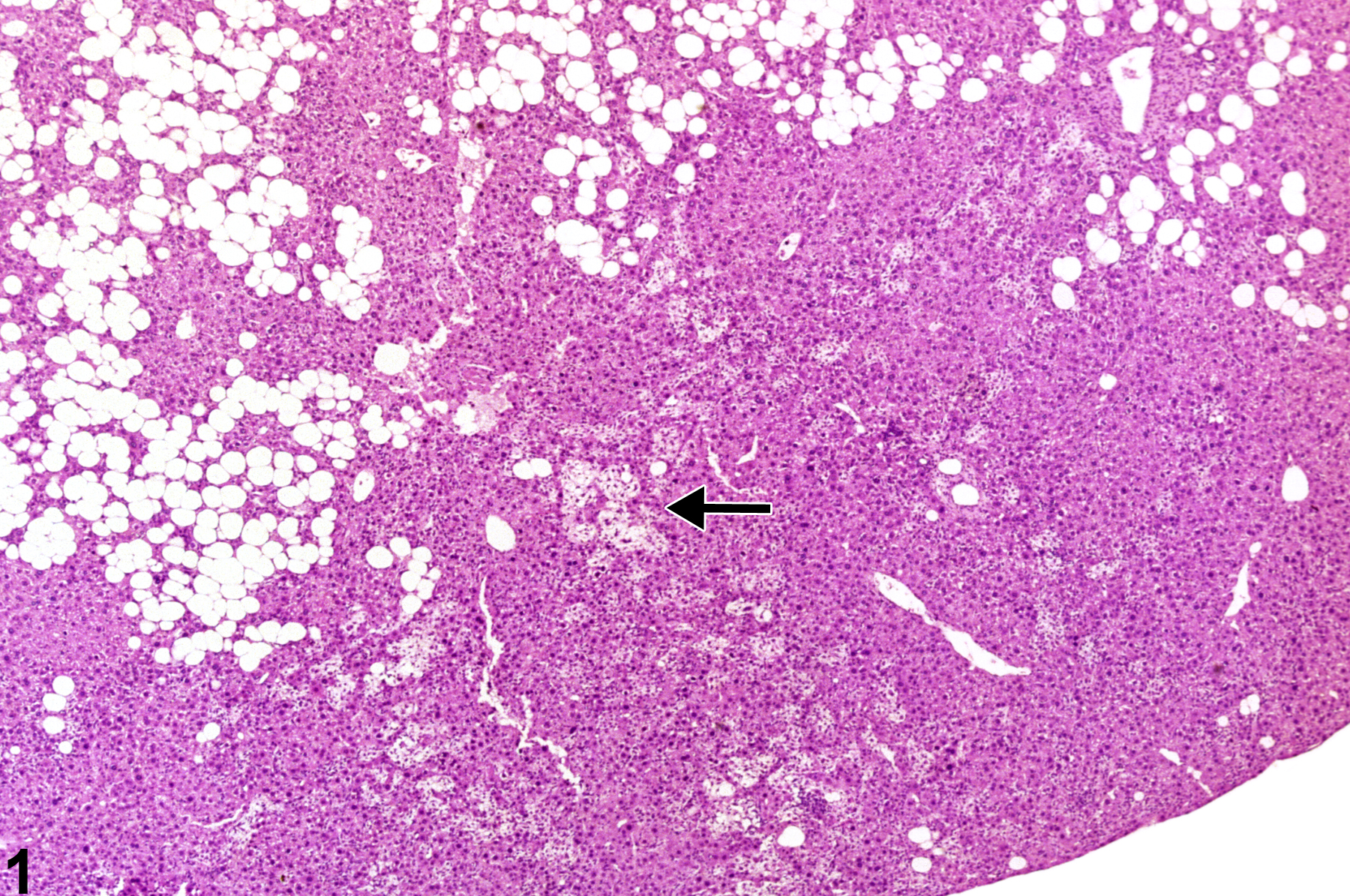
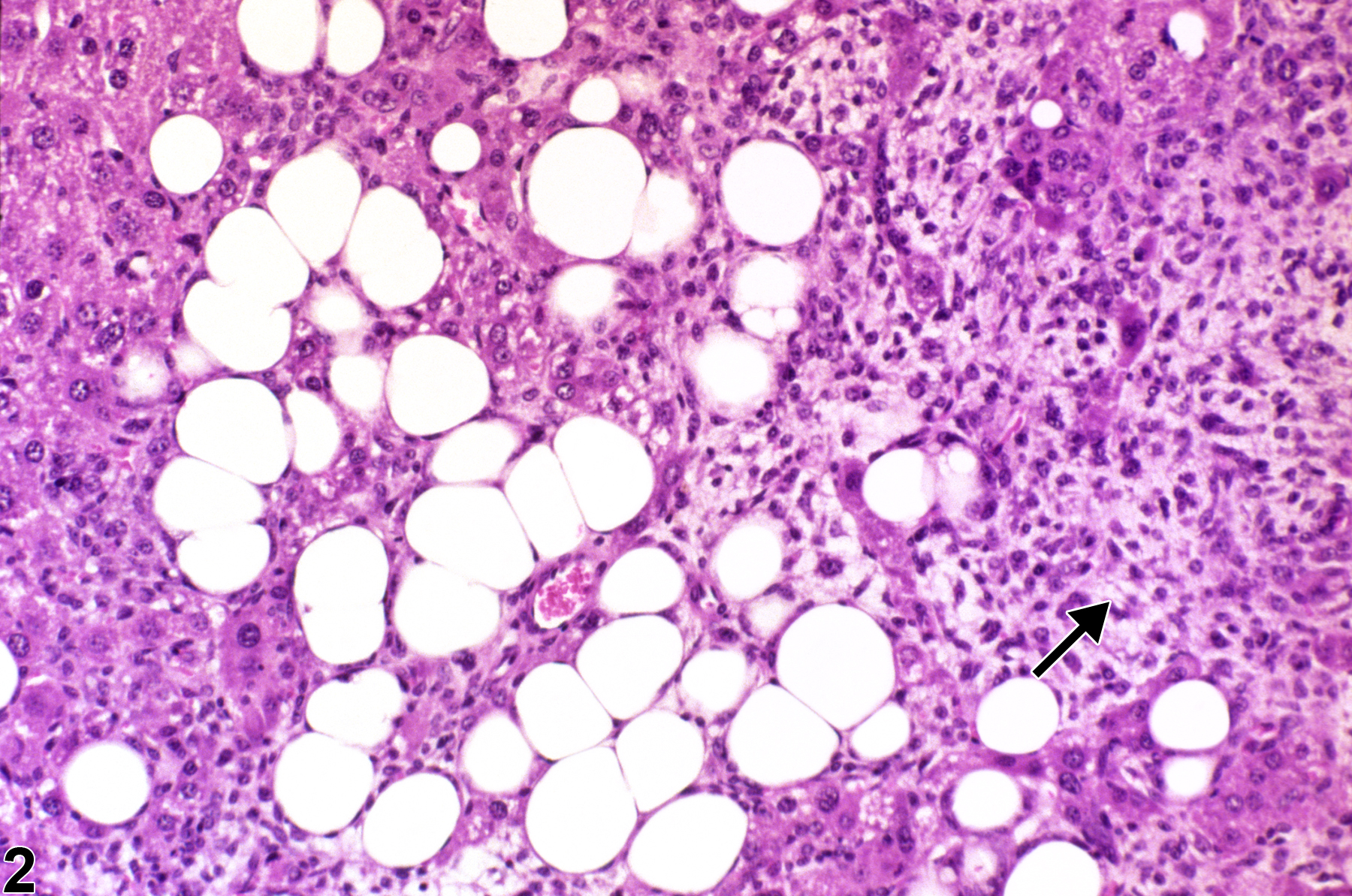
Stellate cell hyperplasia-arrow indicates spindloid morphology in a female B6C3F1 mouse from a chronic study.
Figure 1 of 2
Image of hyperplasia in the liver from a female B6C3F1 mouse in a chronic study
Stellate cell hyperplasia-arrow indicates stellate cells with a spindloid morphology in a female B6C3F1 mouse from a chronic study.
Figure 2 of 2
next arrow

comment:

Hepatic stellate cell (Ito cell or perisinusoidal cell) hyperplasia is rare in mice and has not been documented in NTP rat studies. It consists of a diffuse or multifocal proliferation of stellate cells (fat-storing cells, as opposed to macrovesicular fat within hepatocytes) without prominent compression of surrounding parenchyma. Stellate cells store retinoid compounds and when activated may convert to a spindloid morphology ( Figure 1image opens in a pop-up window and Figure 2image opens in a pop-up window , arrow) and produce extracellular matrix proteins and collagen (scar tissue) in response to hepatic injury. Distinction from stellate cell tumors is based on the latter being more discrete and causing compression of surrounding hepatic parenchyma.

recommendation:

Proliferations of stellate cells should be documented and given a severity grade. If stellate cell tumors are present in the same study, it should be mentioned in the pathology narrative.

references:

Dixon D, Yoshitomi K, Boorman GA, Maronpot RR. 1994. “Lipomatous” lesions of unknown cellular origin in the liver of B6C3F1 mice. Vet Pathol 31:173-182.
Abstract: https://www.ncbi.nlm.nih.gov/pubmed/8203079

Harada T, Enomoto A, Boorman GA, Maronpot RR. 1999. Liver and gallbladder. In: Pathology of the Mouse: Reference and Atlas (Maronpot RR, Boorman GA, Gaul BW, eds). Cache River Press, Vienna, IL, 119-183.
Abstract: http://www.cacheriverpress.com/books/pathmouse.htm

National Toxicology Program. 1987. NTP TR-324. Toxicology and Carcinogenesis Studies of Boric Acid (CAS No. 10043-35-3) in B6C3F1 Mice (Feed Studies). NTP, Research Triangle Park, NC.
Full Text: https://ntp.niehs.nih.gov/ntp/htdocs/lt_rpts/tr324.pdf

Thoolen B, Maronpot RR, Harada T, Nyska A, Rousseaux C, Nolte T, Malarkey D, Kaufmann W, Kutter K, Deschl U, Nakae D, Gregson R, Winlove M, Brix A, Singl B, Belpoggi F, Ward JM. 2010. Hepatobiliary lesion nomenclature and diagnostic criteria for lesions in rats and mice (INHAND). Toxicol Pathol 38:5S-81S.
Full Text: http://tpx.sagepub.com/content/38/7_suppl/5S.full