U.S. flag

An official website of the United States government

Dot gov

The .gov means it's official.
Federal government websites often end in .gov or .mil. Before sharing sensitive information, make sure you're on a federal government site.

Https

The site is secure.
The https:// ensures that you are connecting to the official website and that any information you provide is encrypted and transmitted securely.

Skip to Main Navigation
Skip to Page Content
Skip to Atlas Navigation

Lymph Node - Apoptosis, Lymphocyte

Image of apoptosis, lymphocyte in the lymph node from a female B6C3F1/N mouse in a subchronic study
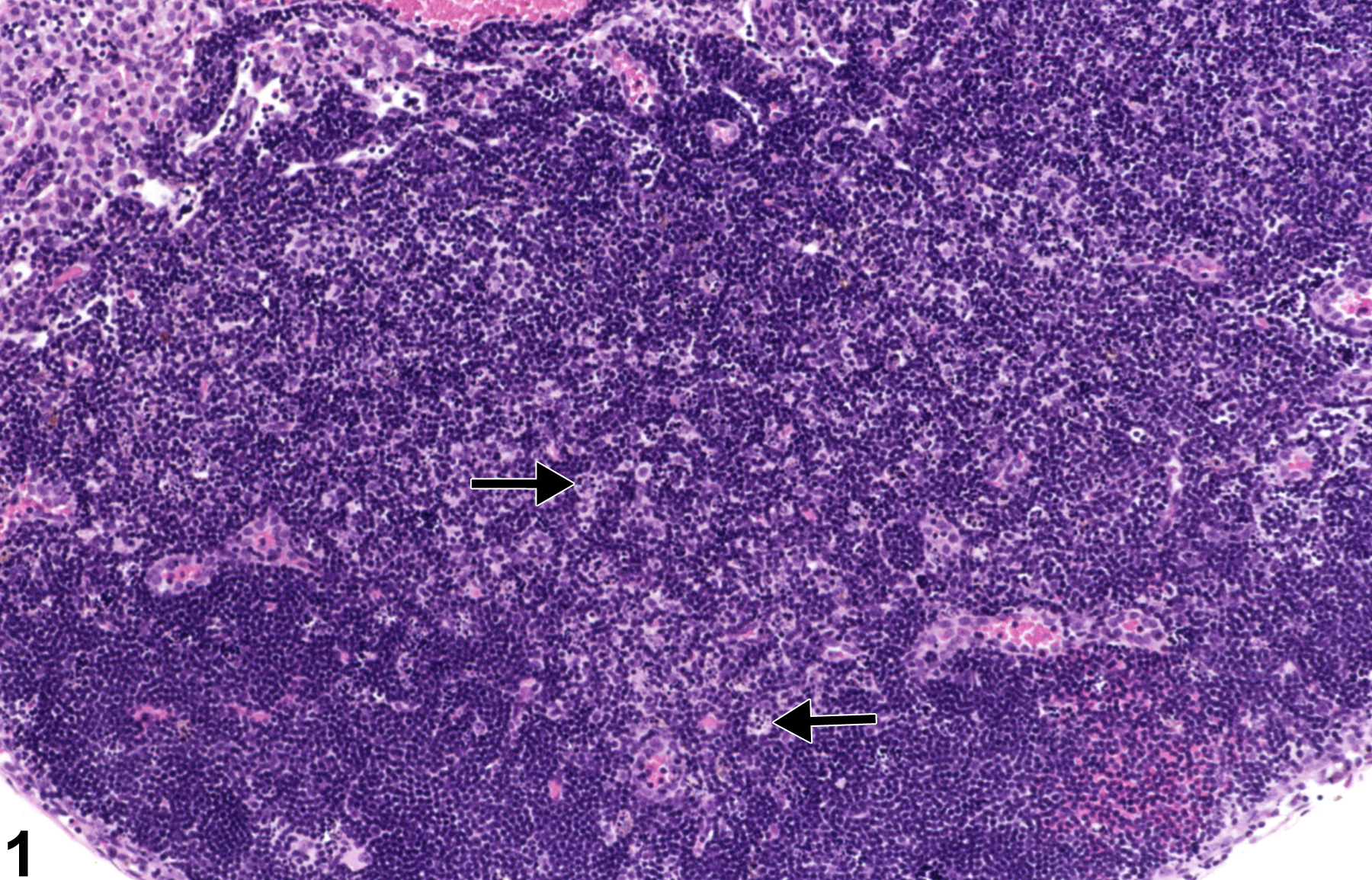
Lymph node - Apoptosis, Lymphocyte in a female B6C3F1/N mouse from a subchronic study. Scattered throughout the paracortex are numerous tingible-body macrophages (arrows).
Figure 1 of 2
Image of apoptosis, lymphocyte in the lymph node from a female B6C3F1/N mouse in a subchronic study
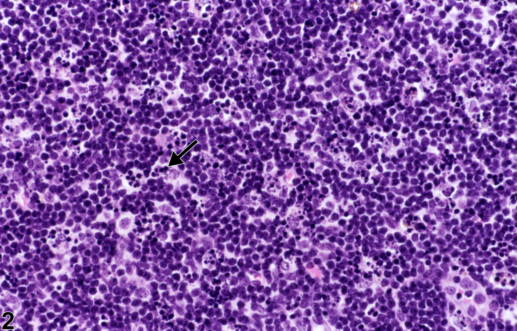
Lymph node - Apoptosis, Lymphocyte in a female B6C3F1/N mouse from a subchronic study (higher magnification of Figure 1). Tingible-body macrophages contain fragments of apoptotic lymphocytes (apoptotic bodies) (arrow).
Figure 2 of 2
next arrow

comment:

Lymphocyte apoptosis is characterized by cell shrinkage, nuclear pyknosis, and fragmentation of lymphocytes into membrane-bound (apoptotic) bodies that are subsequently phagocytized by macrophages (tingible-body macrophages) ( Figure 1image opens in a pop-up window and Figure 2image opens in a pop-up window , arrowheads). This type of cell death normally occurs in B cells within the germinal centers of secondary follicles, where it is an important homeostatic mechanism. Figure 1image opens in a pop-up window and Figure 2image opens in a pop-up window demonstrate the lesion in T cells in the paracortex. Lymphocyte apoptosis may be normal in lymph nodes, increased in nodes of immunodeficient mice, or induced by various injurious stimuli. Some injurious stimuli induce lymphocyte apoptosis at low doses, whereas at higher doses the stimulus leads to necrosis. Examples of such stimuli include heat, irradiation, hypoxia, and cytotoxic cancer drugs (e.g., cyclophosphamide). Dexamethasone can also incite lymphocyte apoptosis; however, the thymus and spleen are more sensitive to this effect than is the lymph node. If severe enough, lymphocyte apoptosis may lead to lymph node atrophy. Historically, lymphocytes apoptosis has been called "lymphocyte necrosis," but the correct terminology for this lesion is "lymphocyte apoptosis." Single-cell necrosis of lymphocytes has not previously been described in the lymph node. Lymphocyte apoptosis must be differentiated from necrosis, although both can occur together. Necrosis generally occurs in groups of cells and is characterized by cell swelling, condensation, fragmentation and dissolution of the nucleus, cell lysis, and accumulation of eosinophilic cytoplasmic and karyorrhectic nuclear debris. Inflammation generally accompanies necrosis because cell lysis releases cellular contents, including proinflammatory mediators, into the interstitial space.

recommendation:

Lymphocyte apoptosis in the lymph node can be normal. If increased compared with concurrent controls, lymphocyte apoptosis should be diagnosed and assigned a severity grade.

references:

Elmore SA. 2006. Enhanced histopathology of the lymph nodes. Toxicol Pathol 34:634-647.
Full Text: https://www.ncbi.nlm.nih.gov/pmc/articles/PMC1783683/

Elmore SA. 2006. Histopathology of the lymph nodes. Toxicol Pathol 34:425-454.
Full Text: https://www.ncbi.nlm.nih.gov/pmc/articles/PMC1892634/

Frith CH, Ward JM, Chandra M, Losco PE. 2000. Non-proliferative lesions of the hematopoietic system in rats. In: Guides for Toxicologic Pathology.TP/ARP/AFIP, Washington, DC.
Full Text: https://www.toxpath.org/docs/SSNDC/HematopoieticNonprolifRat.pdf

National Toxicology Program. 2011. NTP TR-536. Toxicology and Carcinogenesis Studies of Bis(2-chloroethoxy)methane (CAS No. 111-91-1) in Rats and B6C3F1 Mice (Dermal Studies). NTP, Research Triangle Park, NC.
Abstract: https://ntp.niehs.nih.gov/go/34791