

Immune System
Lymph Node - Atrophy
Narrative
Elmore SA. 2006. Enhanced histopathology of the lymph nodes. Toxicol Pathol 34:634-647.
Full Text: https://www.ncbi.nlm.nih.gov/pmc/articles/PMC1783683/Elmore SA. 2006. Histopathology of the lymph nodes. Toxicol Pathol 34:425-454.
Full Text: https://www.ncbi.nlm.nih.gov/pmc/articles/PMC1892634/Frith CH, Ward JM, Chandra M, Losco PE. 2000. Non-proliferative lesions of the hematopoietic system in rats. In: Guides for Toxicologic Pathology.TP/ARP/AFIP, Washington, DC.
Full Text: https://www.toxpath.org/docs/SSNDC/HematopoieticNonprolifRat.pdfNational Toxicology Program. 2004. 13-Week Study (No. C20306) of PCN 66/67 (CAS No. PCNCOMPARISN) in F344 Rats and Sprague-Dawley Rats (Gavage Studies). NTP, Research Triangle Park, NC.
Stefanski SA, Elwell MR, Stromberg PC. 1990. Spleen, lymph nodes, and thymus. In: Pathology of the Fischer Rat: Reference and Atlas (Boorman GA, Eustis SL, Elwell MR, Montgomery CA, MacKenzie WF, eds). Academic Press, San Diego, 369-394.
Ward JM, Mann PC, Morishima H, Frith CH. 1999. Thymus, spleen, and lymph nodes. In: Pathology of the Mouse (Maronpot RR, ed). Cache River Press, Vienna, IL, 333-360.
Lymph node - Atrophy in a female F344/N rat from a subchronic study. With early lymph node atrophy, paracortical lymphocytes are depleted (arrow), and number and size of follicles are decreased (arrowhead).
All Images

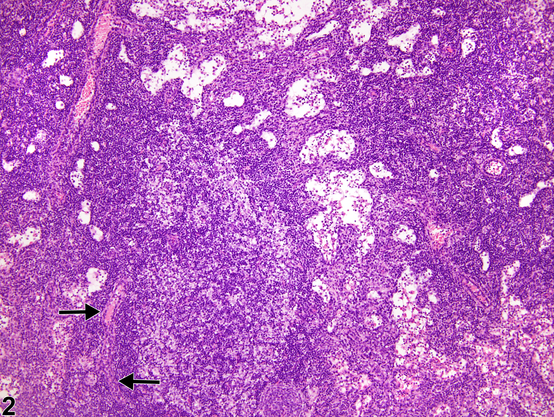
Lymph node - Atrophy in a female F344/N rat from a subchronic study. With early lymph node atrophy, paracortical lymphocytes are depleted (arrow), and number and size of follicles are decreased (arrowhead).

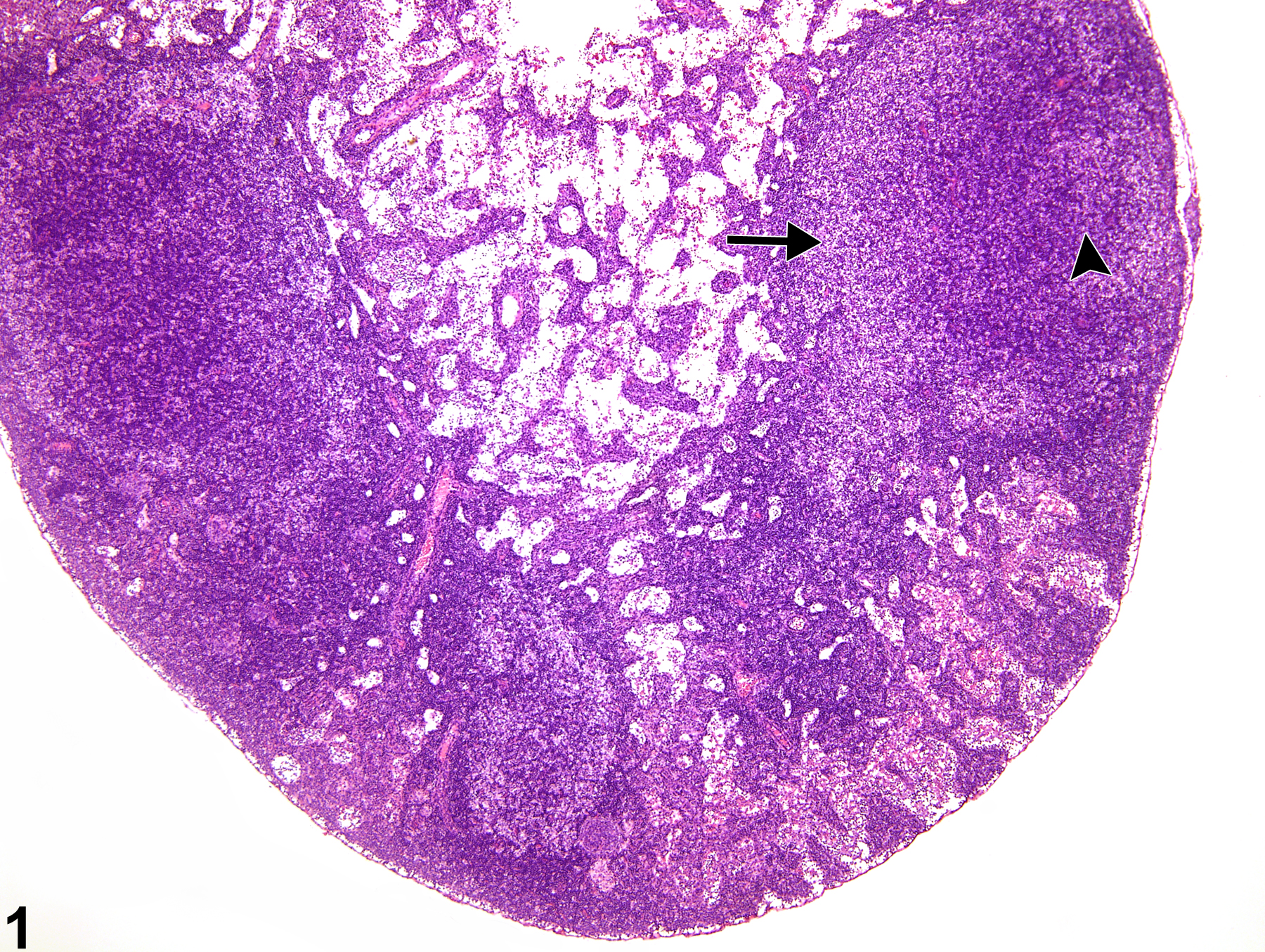
Lymph node - Atrophy in a female F344/N rat from a subchronic study (higher magnification of Figure 1). High endothelial venules (arrows) may be more prominent with lymphocyte depletion.