

Immune System
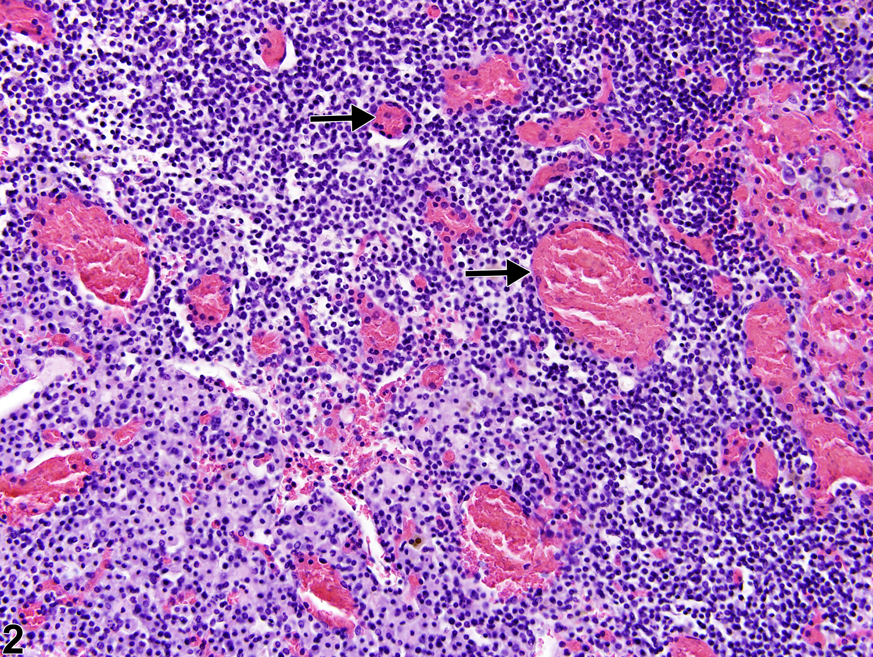
Lymph Node - Congestion
Narrative
Elmore SA. 2006. Histopathology of the lymph nodes. Toxicol Pathol 34:425-454.
Full Text: https://www.ncbi.nlm.nih.gov/pmc/articles/PMC1892634/National Toxicology Program 2010. NTP TR-555. Toxicology and Carcinogenesis Studies of 1,2-Dibromo-2,4-Dicyanobutane (CAS No. 35691-65-7) in F344/N Rats and B6C3F1 Mice (Dermal Studies). NTP, Research Triangle Park, NC.
Abstract: https://ntp.niehs.nih.gov/go/32614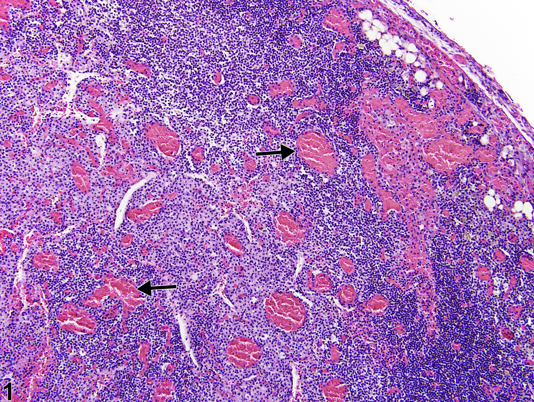
Lymph node - Congestion in a female B6C3F1/N mouse from a chronic study. Numerous vessels are expanded by an excessive accumulation of blood (arrows).
All Images

Lymph node - Congestion in a female B6C3F1/N mouse from a chronic study. Numerous vessels are expanded by an excessive accumulation of blood (arrows).

Lymph node - Congestion in a female B6C3F1/N mouse from a chronic study (higher magnification of Figure 1). Blood vessels contain increased numbers of erythrocytes and other blood elements (arrows).