U.S. flag

An official website of the United States government

Dot gov

The .gov means it's official.
Federal government websites often end in .gov or .mil. Before sharing sensitive information, make sure you're on a federal government site.

Https

The site is secure.
The https:// ensures that you are connecting to the official website and that any information you provide is encrypted and transmitted securely.

Skip to Main Navigation
Skip to Page Content
Skip to Atlas Navigation

Lymph Node - Extramedullary Hematopoiesis

Image of extramedullary hematopoiesis in the lymph node from a female B6C3F1/N mouse in a chronic study
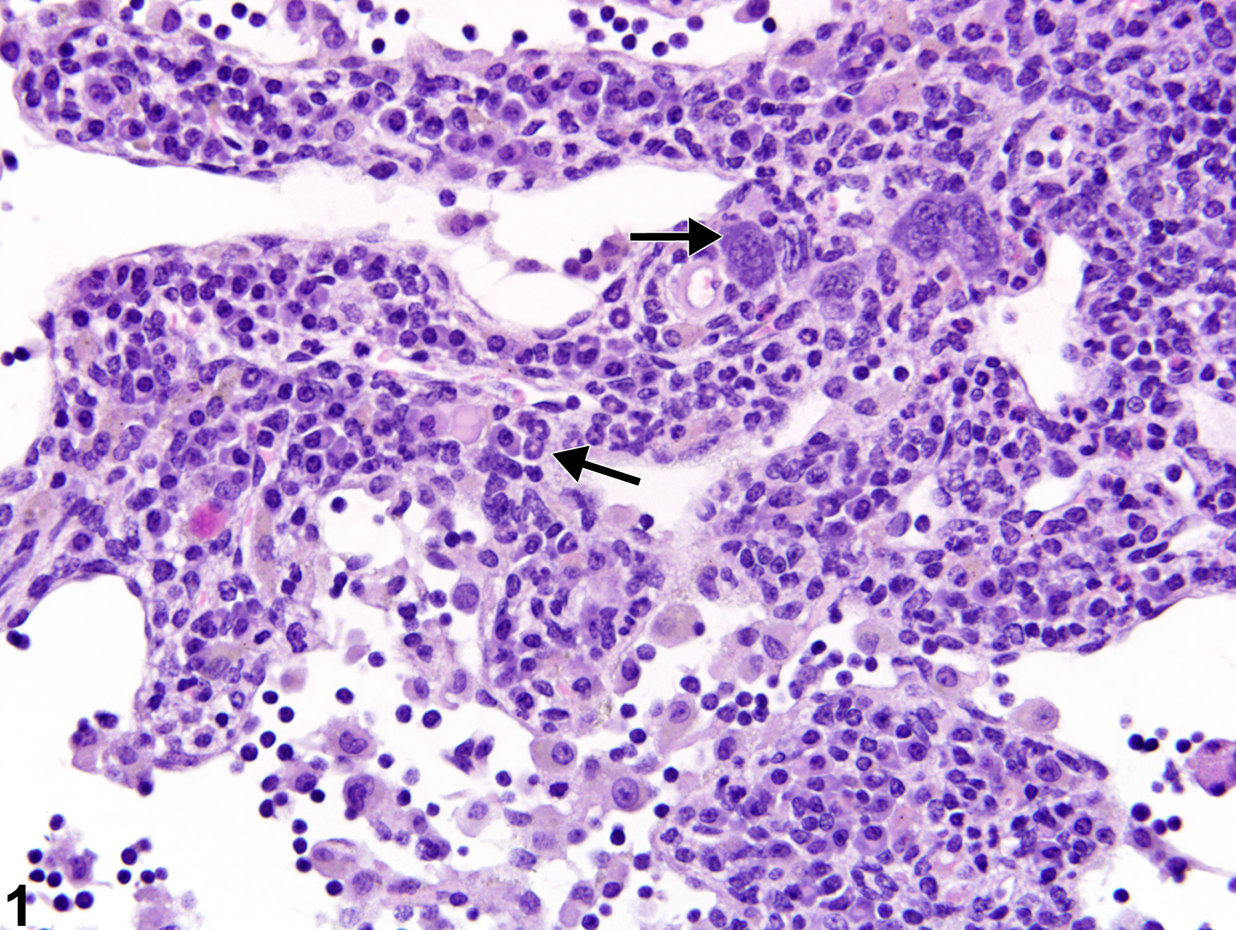
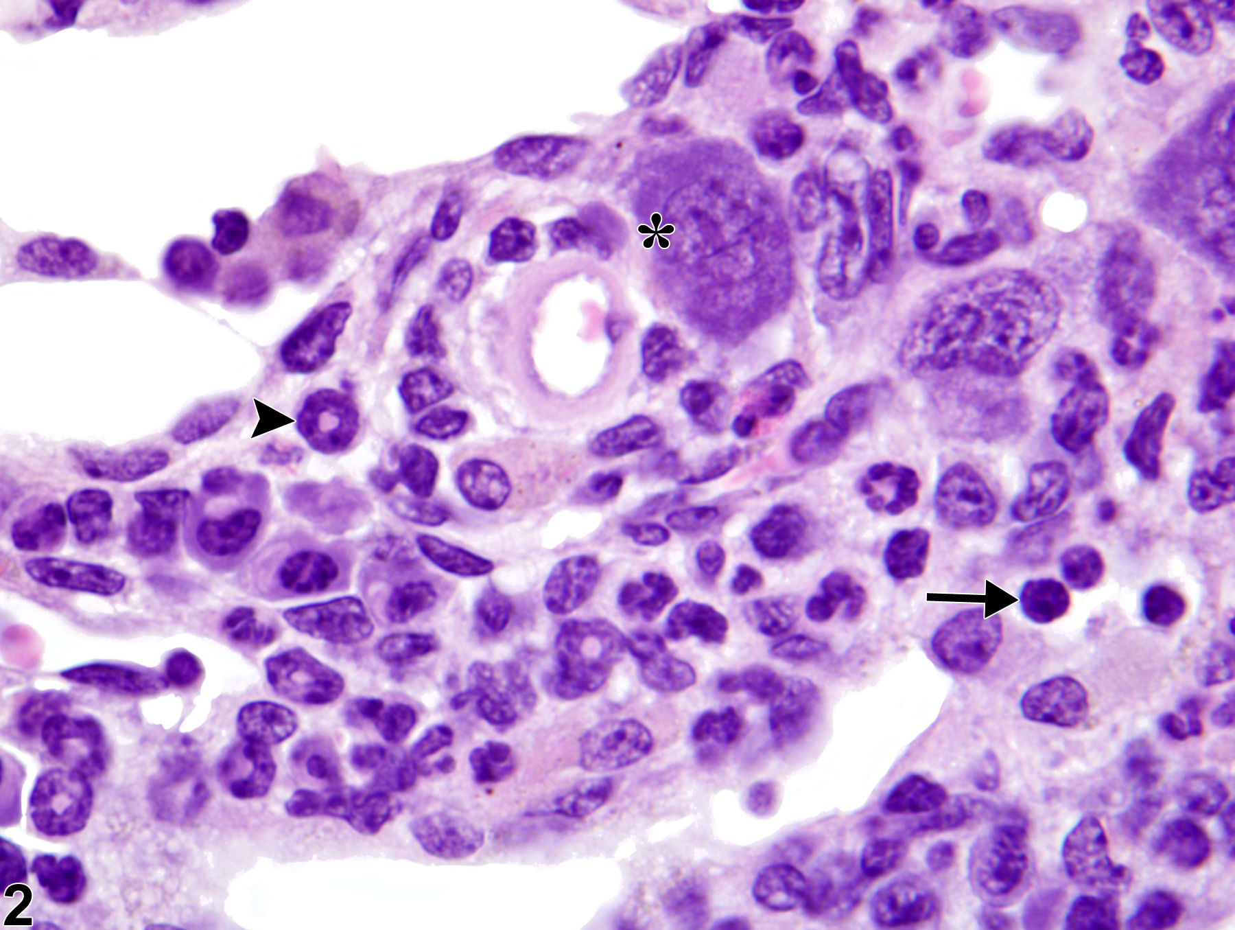
Lymph node - Extramedullary hematopoiesis in a female B6C3F1/N mouse from a chronic study. Increased numbers of hematopoietic cells are present within the lymph node medullary cords (arrows).
Figure 1 of 2
Image of extramedullary hematopoiesis in the lymph node from a female B6C3F1/N mouse in a chronic study
Lymph node - Extramedullary hematopoiesis in a female B6C3F1/N mouse from a chronic study (higher magnification of Figure 1). Hematopoietic cells include erythroblasts (arrow), immature granulocytes (arrowhead), and megakaryocytes (asterisk).
Figure 2 of 2
next arrow

comment:

While the bone marrow is the major site of hematopoiesis, lymph nodes may be a site of extramedullary hematopoiesis (EMH). EMH in lymph nodes typically occurs as a physiologic response to a dramatic loss or increased need for additional blood cells. Examples of inciting factors include hemorrhage, infection, severe inflammation, myelofibrosis, and neoplasia. EMH is characterized by a mixture, to varying degrees, of myeloid, erythroid, and megakaryocytic cells and is primarily present within the lymph node medullary cords ( Figure 1image opens in a pop-up window and Figure 2image opens in a pop-up window ). One lineage of hematopoietic cell may predominate, and various degrees of immaturity may be seen. EMH should be distinguished from inflammatory infiltrates and granulocytic leukemia. Inflammatory infiltrates in the lymph node are generally mature in development and may be a single or mixed cell type (see Lymph Node - Inflammation). Granulocytic leukemia is characterized by high numbers of immature neoplastic myeloid cells that involve multiple organs. Previous terms for extramedullary hematopoiesis include 'hematopoietic cell proliferation" and "myeloid hyperplasia."

recommendation:

Extramedullary hematopoiesis in lymph nodes should be diagnosed and graded. The presence of this lesion in lymph nodes should prompt evaluation of other organs for EMH (especially the spleen and liver).

references:

Dunn TB. 1954. Normal and pathologic anatomy of the reticular tissue in laboratory mice. J Nat Cancer Inst 14:1281-1433.
Abstract: http://jnci.oxfordjournals.org/content/14/6/1281.extract

Elmore SA. 2006. Histopathology of the lymph nodes. Toxicol Pathol 34:425-454.
Full Text: https://www.ncbi.nlm.nih.gov/pmc/articles/PMC1892634/

Elmore SA. 2006. Enhanced histopathology of the lymph nodes. Toxicol Pathol 34:634-647.
Full Text: https://www.ncbi.nlm.nih.gov/pmc/articles/PMC1783683/

Kim CH. 2010. Homeostatic and pathogenic extramedullary hematopoiesis. J Blood Med 1:13-19.
Full Text: https://www.ncbi.nlm.nih.gov/pmc/articles/PMC3262334/

National Toxicology Program. 2011. NTP TR-570. Toxicology and Carcinogenesis Studies of α,β-Thujone (CAS No. 76231-76-0) in F344/N Rats and B6C3F1 Mice (Gavage Studies). NTP, Research Triangle Park, NC.
Abstract: https://ntp.niehs.nih.gov/go/36137