U.S. flag

An official website of the United States government

Dot gov

The .gov means it's official.
Federal government websites often end in .gov or .mil. Before sharing sensitive information, make sure you're on a federal government site.

Https

The site is secure.
The https:// ensures that you are connecting to the official website and that any information you provide is encrypted and transmitted securely.

Skip to Main Navigation
Skip to Page Content
Skip to Atlas Navigation

Lymph Node - Hyperplasia, Plasma Cell
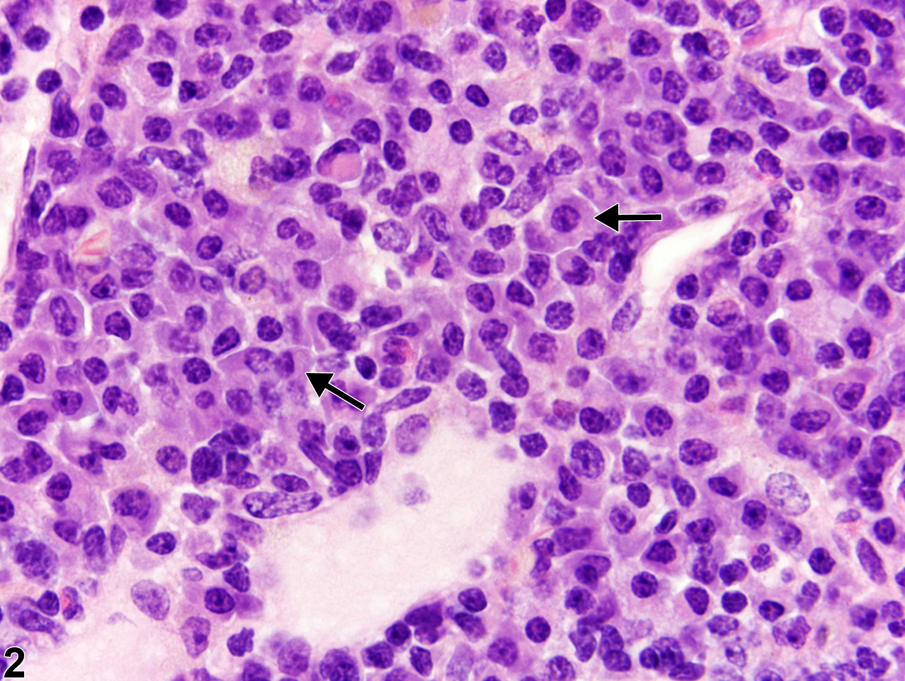
Image of hyperplasia, plasma cell in the lymph node from a female B6C3F1/N mouse in a chronic study
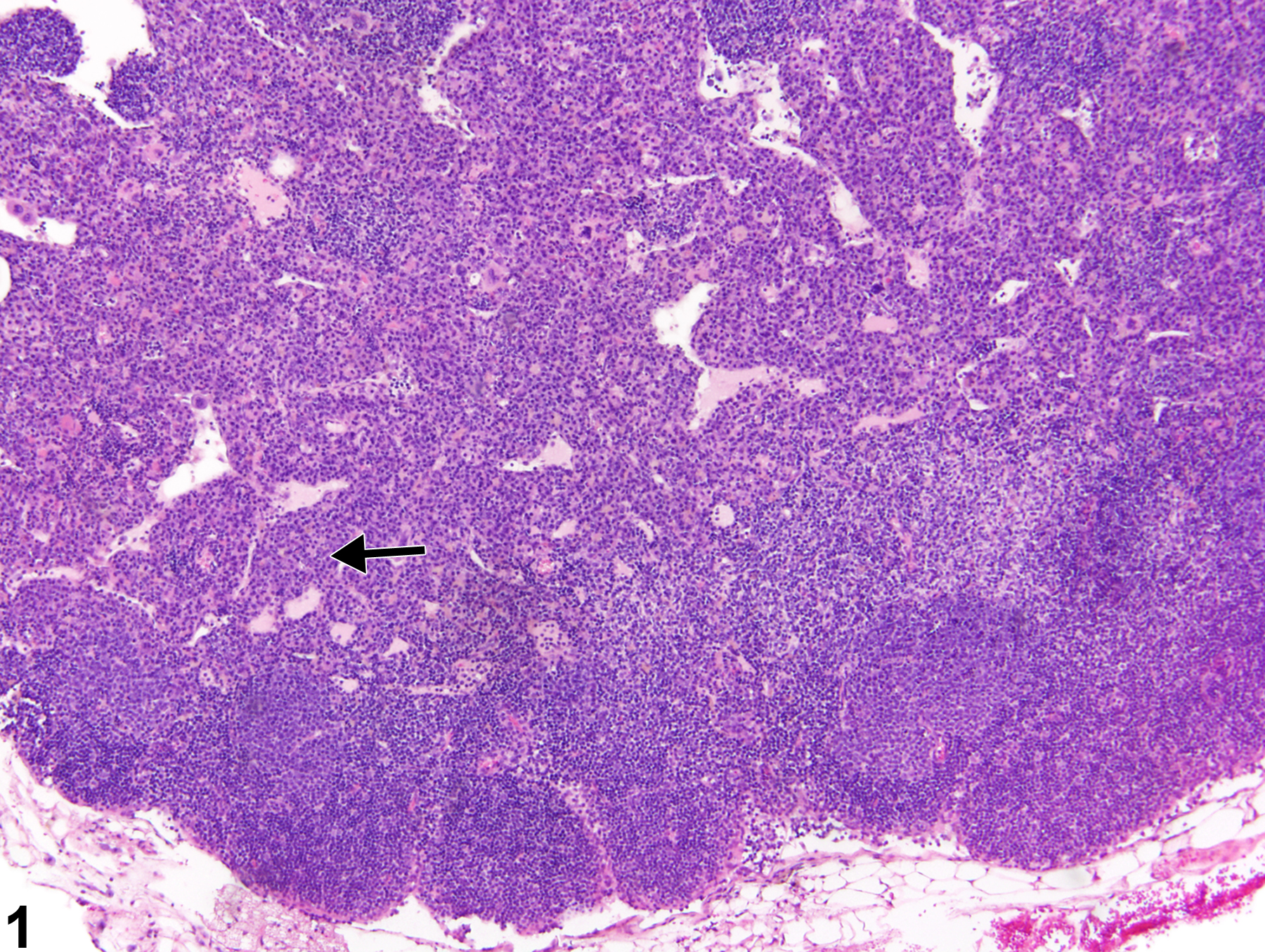
Lymph node - Hyperplasia, Plasma cell in a control female B6C3F1/N mouse from a chronic study. The medullary cords are markedly expanded by increased numbers of plasma cells (arrow).
Figure 1 of 2
Image of hyperplasia, plasma cell in the lymph node from a female B6C3F1/N mouse in a chronic study
Lymph node - Hyperplasia, Plasma cell in a control female B6C3F1/N mouse from a chronic study (higher magnification of Figure 1). Plasma cells (arrows) are present within the medullary cords.
Figure 2 of 2
next arrow

comment:

Plasma cell hyperplasia is a common finding in the lymph nodes of rodents, particularly in the mandibular lymph nodes ( Figure 1image opens in a pop-up window and Figure 2image opens in a pop-up window ), most often in response to exposure to dietary antigens. If not a background lesion, then plasma cell hyperplasia may be treatment related or associated with inflammatory, infectious, or neoplastic lesions in other organs. Plasma cells and their precursors are normally present in the medullary cords; therefore, these cords are the primary site of plasma cell hyperplasia ( Figure 1image opens in a pop-up window ). Plasma cells usually increase in response to antigenic stimulation requiring antibody production, so typically B-cell (lymphoid) hyperplasia is accompanied by plasma cell hyperplasia. Myeloid hyperplasia may also be present. Mature plasma cells have basophilic to eosinophilic cytoplasm, eccentrically located nuclei, a Golgi clear zone, and a cartwheel chromatin pattern ( Figure 2image opens in a pop-up window , arrows). Depending on the degree and chronicity of antigenic stimulation, some Mott cells with Russell bodies may be present. With marked plasma cell hyperplasia, the lymph node may be greatly enlarged and composed mostly of plasma cells and may exhibit partial effacement of normal nodal architecture. Marked plasma cell hyperplasia may be differentiated from plasma cell neoplasia by the presence of plasma cell precursors, atypical plasma cells, capsular infiltration, and metastases in the latter.

recommendation:

Plasma cell hyperplasia in lymph nodes should be diagnosed and given a severity grade. However, if this lesion accompanies lymphocyte hyperplasia and/or neoplasia, it should not be diagnosed separately but should be described in the narrative.

references:

Elmore SA. 2006. Enhanced histopathology of the lymph nodes. Toxicol Pathol 34:634-647.
Full Text: https://www.ncbi.nlm.nih.gov/pmc/articles/PMC1783683/

Elmore SA. 2006. Histopathology of the lymph nodes. Toxicol Pathol 34:425-454.
Full Text: https://www.ncbi.nlm.nih.gov/pmc/articles/PMC1892634/

Frith CH, Ward JM, Chandra M, Losco PE. 2000. Non-proliferative lesions of the hematopoietic system in rats. In: Guides for Toxicologic Pathology.TP/ARP/AFIP, Washington, DC.
Full Text: https://www.toxpath.org/docs/SSNDC/HematopoieticNonprolifRat.pdf

National Toxicology Program. 2004. NTP TR-511. Toxicology and Carcinogenesis Studies of Dipropylene Glycol (CAS No. 25265-71-8) in F344/N Rats and B6C3F1 Mice (Drinking Water Studies). NTP, Research Triangle Park, NC.
Abstract: https://ntp.niehs.nih.gov/go/14900

Ward JM, Mann PC, Morishima H, Frith CH. 1999. Thymus, spleen, and lymph nodes. In: Pathology of the Mouse (Maronpot RR, ed). Cache River Press, Vienna, IL, 333-360.

Ward JM, Rehg JE, Morse HC III. 2012. Differentiation of rodent immune and hematopoietic system reactive lesions from neoplasia. Toxicol Pathol 40:425-434.
Abstract: https://www.ncbi.nlm.nih.gov/pubmed/22215512