

Immune System
Spleen - Angiectasis
Narrative
Elmore SA. 2006. Histopathology of the lymph nodes. Toxicol Pathol 34:425-454.
Full Text: https://www.ncbi.nlm.nih.gov/pmc/articles/PMC1892634/National Toxicology Program. 2010. NTP TR-556. Toxicology and Carcinogenesis Studies of Chromium Picolinate Monohydrate (CAS No. 27882-76-4) in F344/N Rats and B6C3F1 Mice (Feed Studies). NTP, Research Triangle Park, NC.
Abstract: https://ntp.niehs.nih.gov/go/32608National Toxicology Program. 2011. NTP TR-570. Toxicology and Carcinogenesis Studies of α,β-Thujone (CAS No. 76231-76-0) in F344/N Rats and B6C3F1 Mice (Gavage Studies). NTP, Research Triangle Park, NC.
Abstract: https://ntp.niehs.nih.gov/go/36137Stefanski SA, Elwell MR, Stromberg PC. 1990. Spleen, lymph nodes, and thymus. In: Pathology of the Fischer Rat: Reference and Atlas (Boorman GA, Eustis SL, Elwell MR, Montgomery CA, MacKenzie WF, eds). Academic Press, San Diego, 369-394.
Ward JM, Mann PC, Morishima H, Frith CH. 1999. Thymus, spleen, and lymph nodes. In: Pathology of the Mouse (Maronpot RR, ed). Cache River Press, Vienna, IL, 333-360.
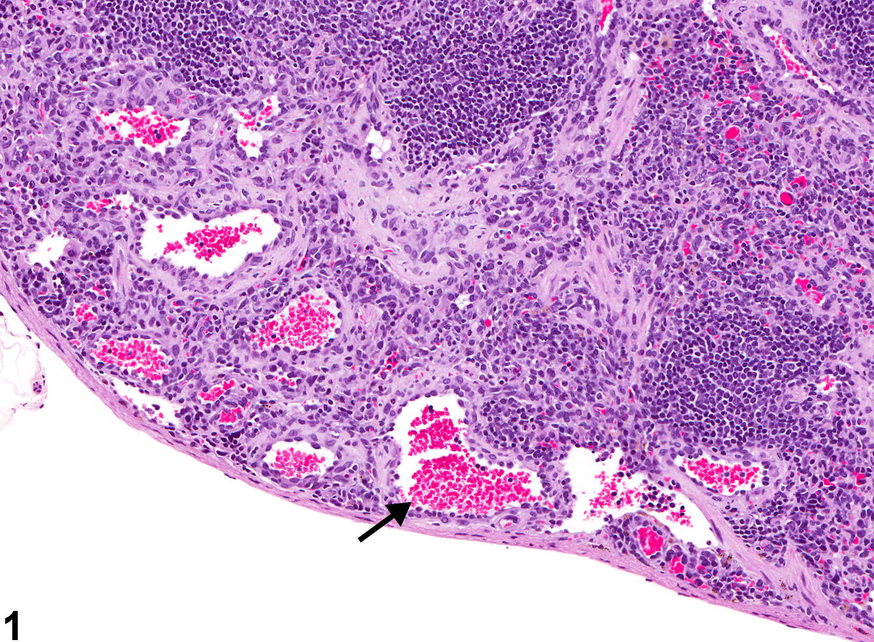
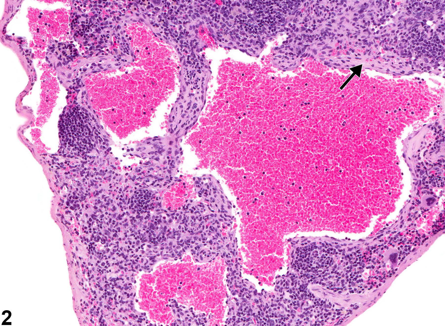
Spleen - Angiectasis in a female B6C3F1/N mouse from a chronic study. Multiple variably sized blood-filled spaces lined by endothelium (arrow) are present within the splenic parenchyma.
All Images

Spleen - Angiectasis in a female B6C3F1/N mouse from a chronic study. Multiple variably sized blood-filled spaces lined by endothelium (arrow) are present within the splenic parenchyma.

Spleen - Angiectasis in a female B6C3F1/N mouse from a chronic study. Angiectasis is associated with fibrosis (arrow) in the parenchyma of this spleen.