U.S. flag

An official website of the United States government

Dot gov

The .gov means it's official.
Federal government websites often end in .gov or .mil. Before sharing sensitive information, make sure you're on a federal government site.

Https

The site is secure.
The https:// ensures that you are connecting to the official website and that any information you provide is encrypted and transmitted securely.

Skip to Main Navigation
Skip to Page Content
Skip to Atlas Navigation

Spleen - Apoptosis, Lymphocyte

Image of apoptosis, lymphocyte in the spleen from a female B6C3F1/N mouse in a subchronic study
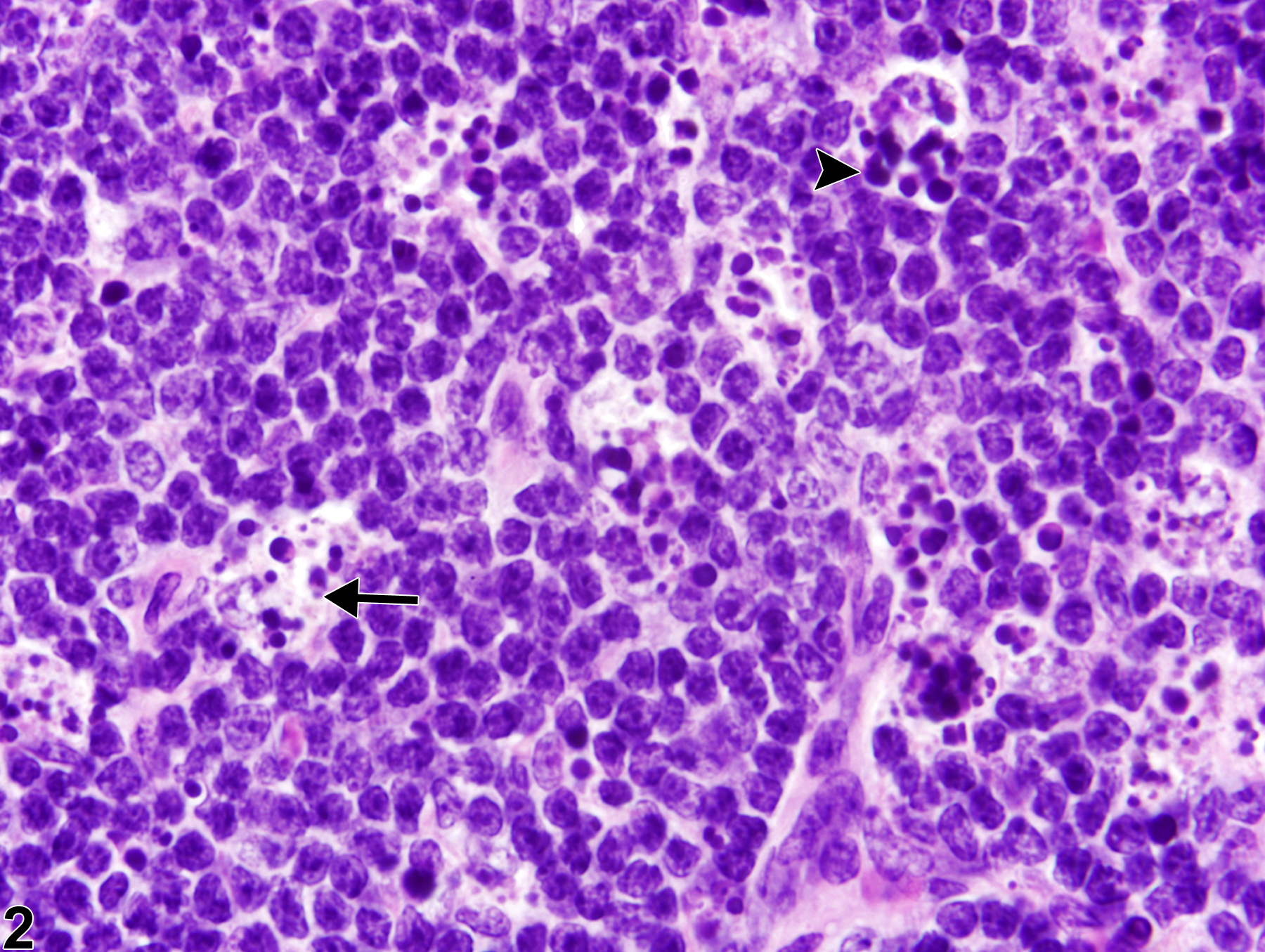
Spleen - Apoptosis, Lymphocyte in a female B6C3F1/N mouse from a subchronic study. Tingible body macrophages are scattered throughout the splenic white pulp (arrows).
Figure 1 of 2
Image of apoptosis, lymphocyte in the spleen from a female B6C3F1/N mouse in a subchronic study
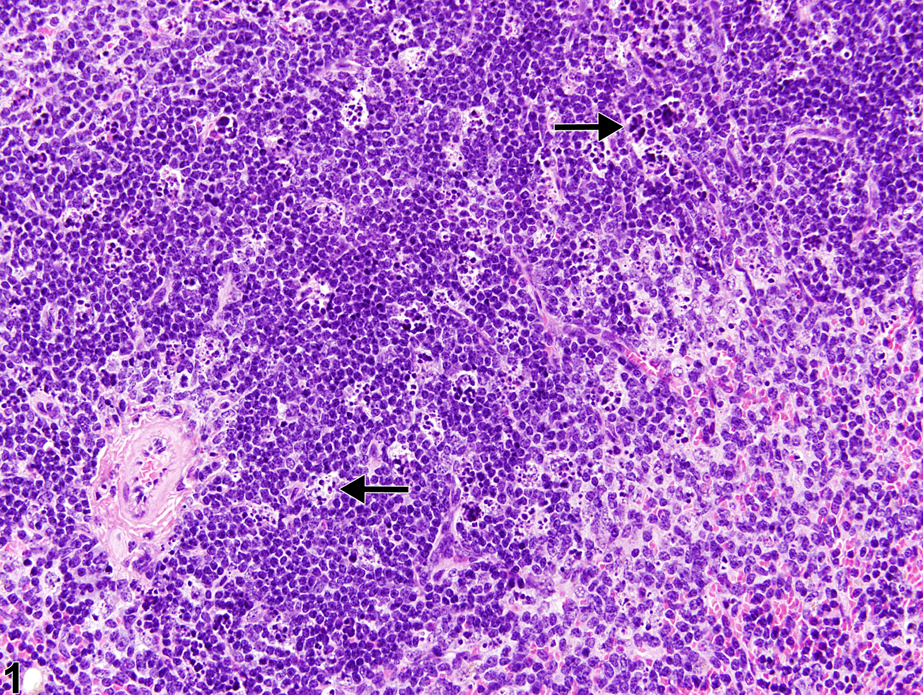
Spleen - Apoptosis, Lymphocyte in a female B6C3F1/N mouse from a subchronic study (higher magnification of Figure 1). Tingible body macrophages (arrow) contain intracytoplasmic fragments of apoptotic lymphocytes (apoptotic bodies) (arrowhead).
Figure 2 of 2
next arrow

comment:

Lymphocyte apoptosis normally occurs in the B-cell-rich follicular germinal centers of the spleen of rodents but may also be increased in B-cell and/or T-cell (periarteriolar lymphatic sheaths) compartments with experimental exposures to radiation, viruses, endotoxin, or chemicals. Apoptosis is characterized by shrinkage of individual lymphocytes, condensation of nuclear chromatin, and fragmentation of apoptotic cells into membrane-bound bodies (apoptotic bodies) ( Figure 2image opens in a pop-up window , arrowhead), which are subsequently phagocytized by macrophages (tingible body macrophages) ( Figure 1image opens in a pop-up window and Figure 2image opens in a pop-up window , arrows). Significant lymphocyte apoptosis may lead to atrophy of one or more white pulp compartments. Historically, apoptosis of lymphocytes has been called "lymphocyte necrosis," but the correct terminology for this lesion is "lymphocyte apoptosis." A diagnosis of necrosis should be used when the predominant cytomorphology is consistent with the classic forms of necrosis (see Spleen - Necrosis). Necrosis is characterized by cell swelling, condensation, fragmentation and dissolution of the nucleus, cell lysis, and accumulation of abundant eosinophilic cytoplasmic and karyorrhectic nuclear debris. Inflammation is generally present with necrosis.

recommendation:

When increased above that seen in concurrent controls, lymphocyte apoptosis in the spleen should be diagnosed and assigned a severity grade. Lymphocyte apoptosis must be differentiated from splenic necrosis.

related links:

Spleen - Necrosis

references:

Elmore SA. 2006. Enhanced histopathology of the spleen. Toxicol Pathol 34:648-655.
Full Text: https://www.ncbi.nlm.nih.gov/pmc/articles/PMC1828535/

National Toxicology Program. 2011. NTP TR-536. Bis(2-chloroethoxy)methane (CAS No. 111-91-1) in F344/N Rats and B6C3F1 Mice (Dermal Studies). NTP, Research Triangle Park, NC.
Abstract: https://ntp.niehs.nih.gov/go/34791

Stefanski SA, Elwell MR, Stromberg PC. 1990. Spleen, lymph nodes, and thymus. In: Pathology of the Fischer Rat: Reference and Atlas (Boorman GA, Eustis SL, Elwell MR, Montgomery CA, MacKenzie WF, eds). Academic Press, San Diego, 369-394.

Suttie AW. 2006. Histopathology of the spleen. Toxicol Pathol 34:466-503.
Full Text: http://tpx.sagepub.com/content/34/5/466.full.pdf

Ward JM, Mann PC, Morishima H, Frith CH. 1999. Thymus, spleen, and lymph nodes. In: Pathology of the Mouse (Maronpot RR, ed). Cache River Press, Vienna, IL, 333-360.