

Immune System
Spleen - Fibrosis
Narrative
Goodman DG, Ward JM, Reichardt WD. 1984. Splenic fibrosis and sarcomas in F344 rats fed diets containing aniline hydrochloride, p-chloroaniline, azobenzene, o-toluidine hydrochloride, 4,4'-sulfonyldianiline, or D&C red no. 9. J Natl Cancer Inst 73:265-273.
Abstract: https://www.ncbi.nlm.nih.gov/pubmed/6588231National Toxicology Program. 2000. NTP TR-484. Toxicology and Carcinogenesis Studies of 2-Butoxyethanol (CAS No. 111-76-2) in F344/N Rats and B6C3F1 Mice (Inhalation Studies). NTP, Research Triangle Park, NC.
Abstract: https://ntp.niehs.nih.gov/go/9770National Toxicology Program. 2008. NTP TR-540. Toxicology and Carcinogenesis Studies of Methylene Blue Trihydrate (CAS No. 7220-79-3) in F344/N Rats and B6C3F1 Mice (Gavage Studies). NTP, Research Triangle Park, NC.
Abstract: https://ntp.niehs.nih.gov/go/32989Stefanski SA, Elwell MR, Stromberg PC. 1990. Spleen, lymph nodes, and thymus. In: Pathology of the Fischer Rat: Reference and Atlas (Boorman GA, Eustis SL, Elwell MR, Montgomery CA, MacKenzie WF, eds). Academic Press, San Diego, 369-394.
Suttie AW. 2006. Histopathology of the spleen. Toxicol Pathol 34:466-503.
Full Text: http://tpx.sagepub.com/content/34/5/466.full.pdfWard JM, Mann PC, Morishima H, Frith CH. 1999. Thymus, spleen, and lymph nodes. In: Pathology of the Mouse (Maronpot RR, ed). Cache River Press, Vienna, IL, 333-360.
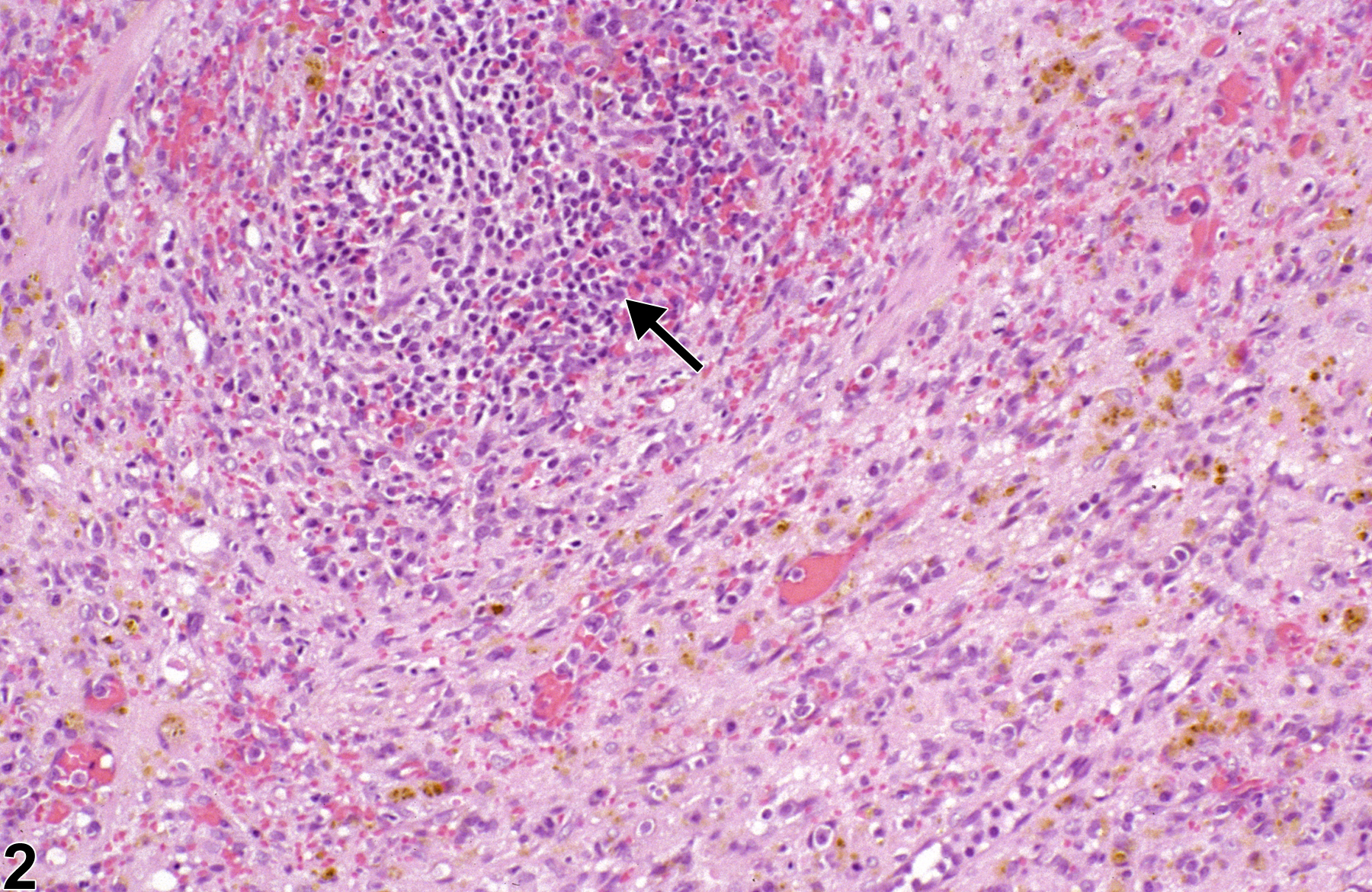
Spleen, Parenchyma - Fibrosis in a male F344/N rat from a chronic study. The splenic red pulp is contracted by fibrous connective tissue (arrow).
All Images

Spleen, Parenchyma - Fibrosis in a male F344/N rat from a chronic study. The splenic red pulp is contracted by fibrous connective tissue (arrow).

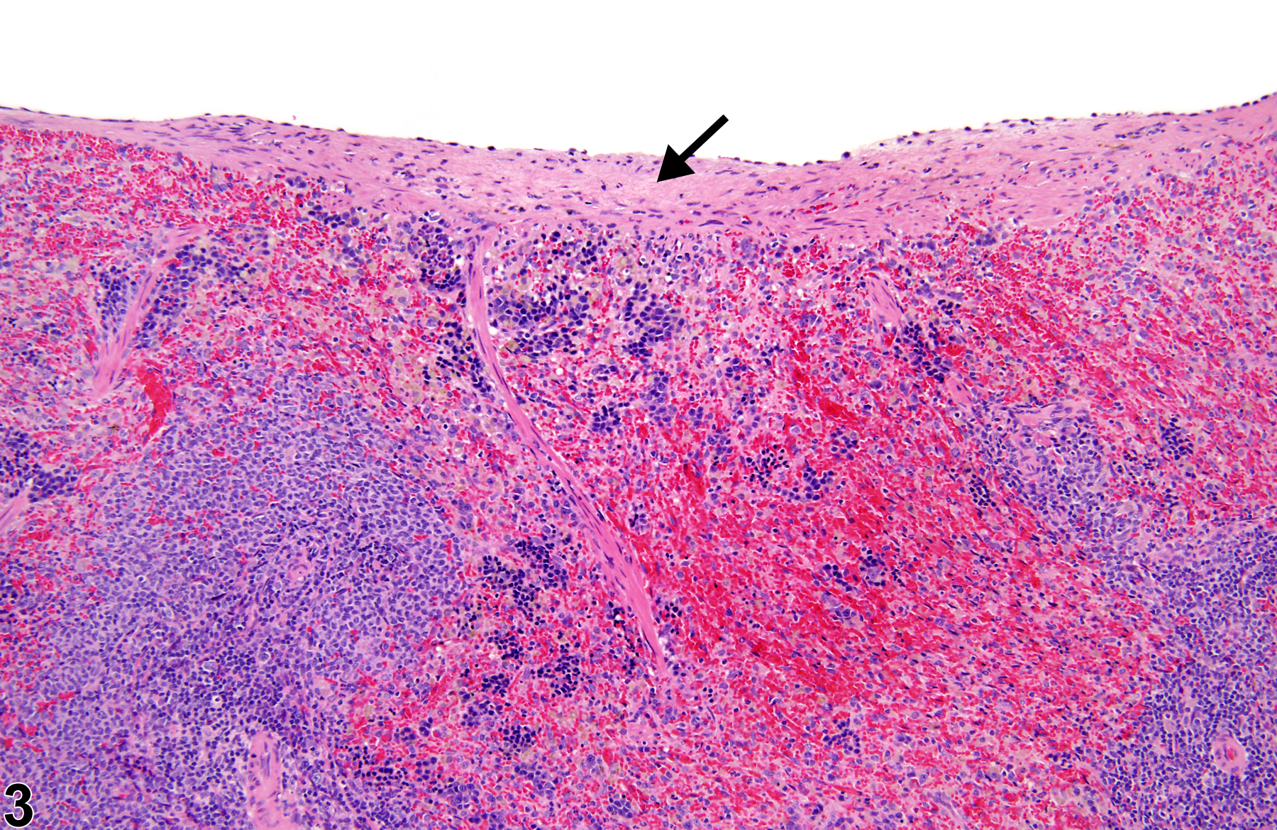
Spleen, Parenchyma - Fibrosis in a male F344/N rat from a chronic study (higher magnification of Figure 1). The splenic white pulp is atrophied (arrow), and there is pigment within the surrounding fibrous connective tissue.

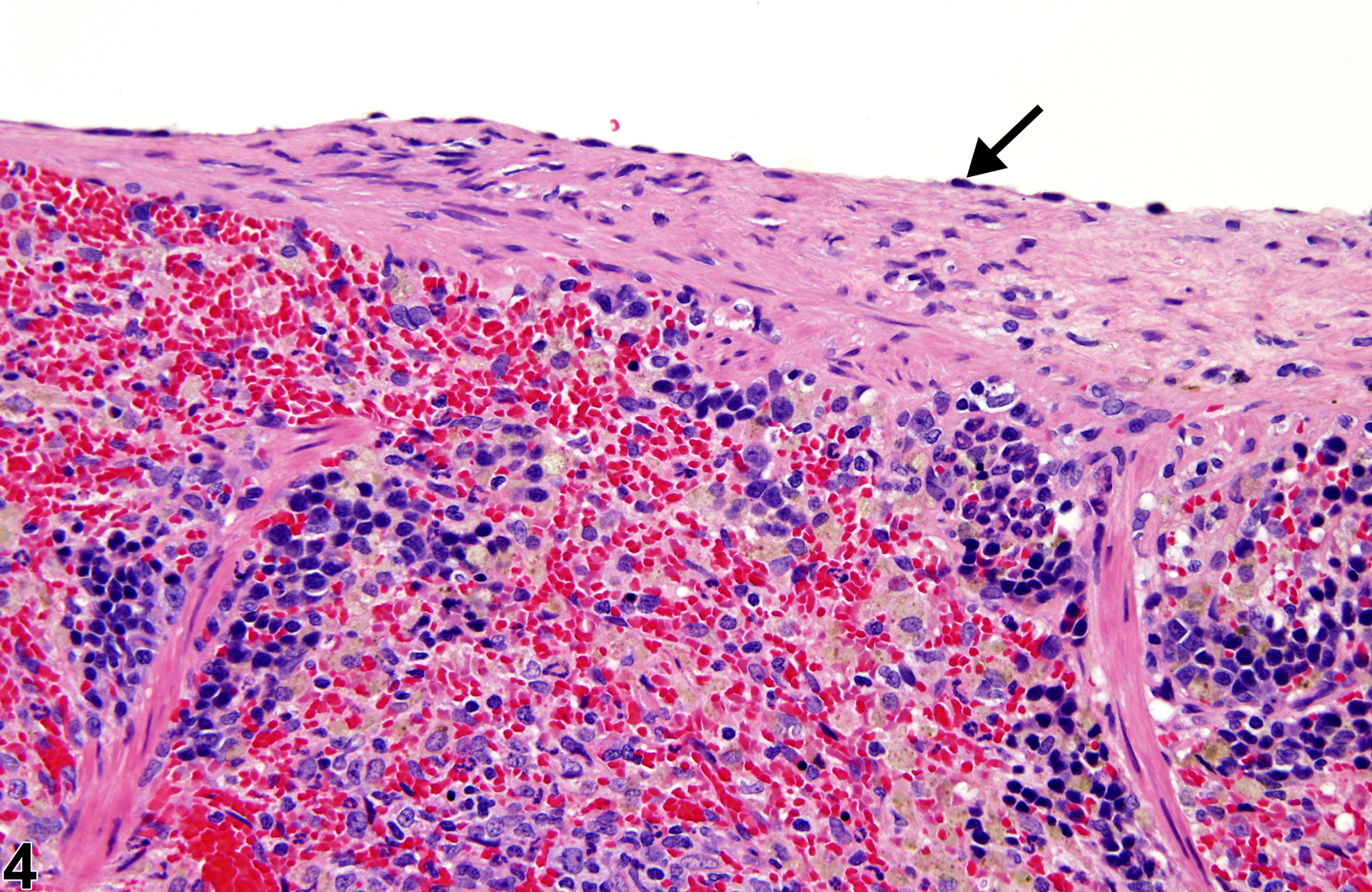
Spleen, Capsule - Fibrosis in a male F344/N rat from a chronic study. The splenic capsule is expanded by fibrous connective tissue (arrow).

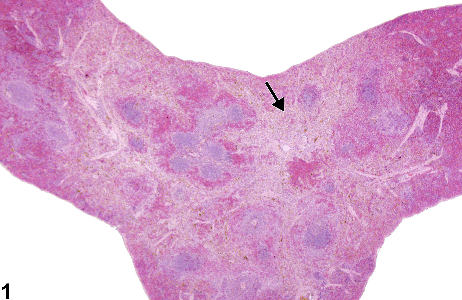
Spleen, Capsule - Fibrosis in a male F344/N rat from a chronic study (higher magnification of Figure 3). The fibrotic splenic capsule is lined by hypertrophied mesothelial cells (arrow).