U.S. flag

An official website of the United States government

Dot gov

The .gov means it's official.
Federal government websites often end in .gov or .mil. Before sharing sensitive information, make sure you're on a federal government site.

Https

The site is secure.
The https:// ensures that you are connecting to the official website and that any information you provide is encrypted and transmitted securely.

Skip to Main Navigation
Skip to Page Content
Skip to Atlas Navigation

Spleen - Hemorrhage

Image of hemorrhage in the spleen from a female Harlan Sprague-Dawley rat in a chronic study
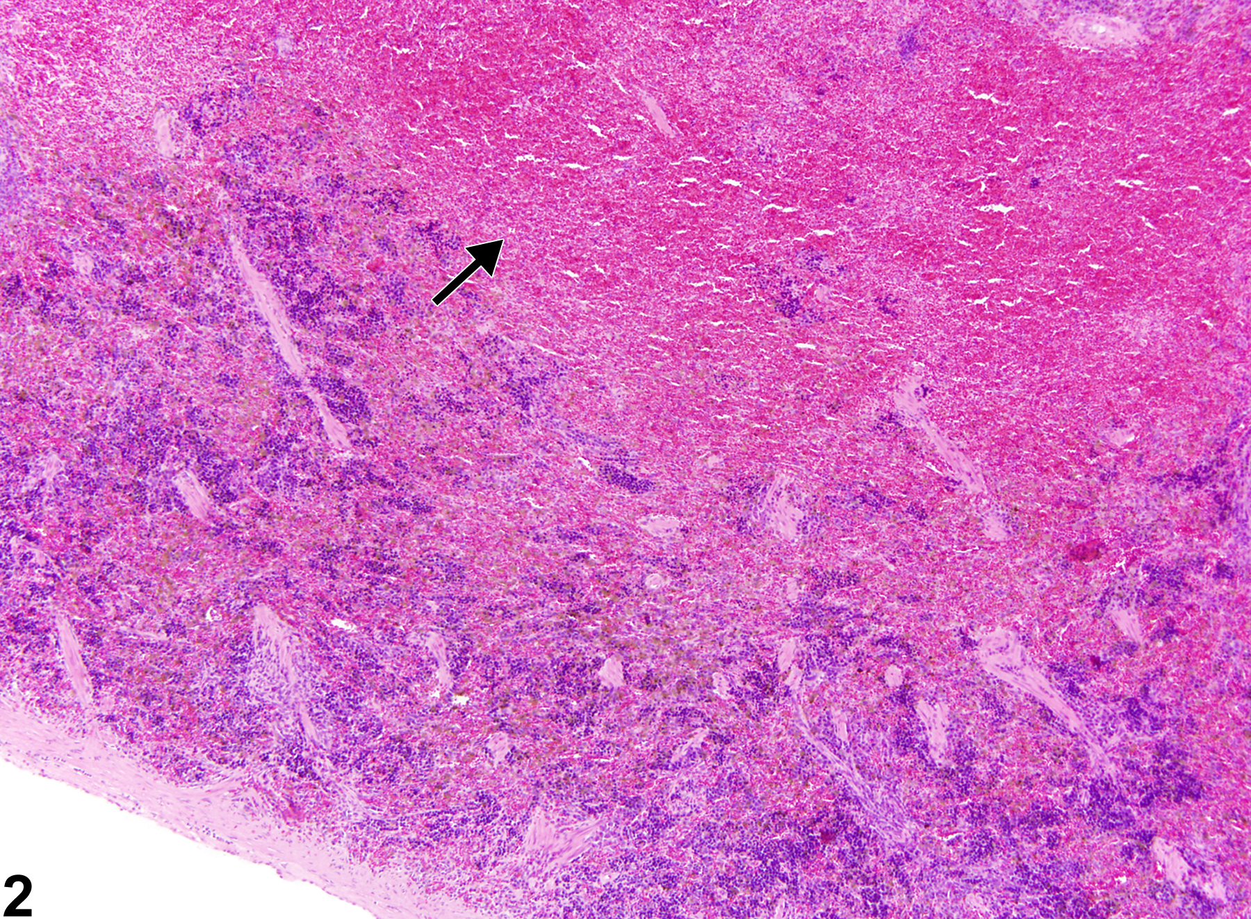
Spleen - Hemorrhage in a female Harlan Sprague-Dawley rat from a chronic study. A focal accumulation of extravasated erythrocytes resulted in protrusion of the splenic capsule (arrow).
Figure 1 of 2
Image of hemorrhage in the spleen from a female F344/N rat in a chronic study
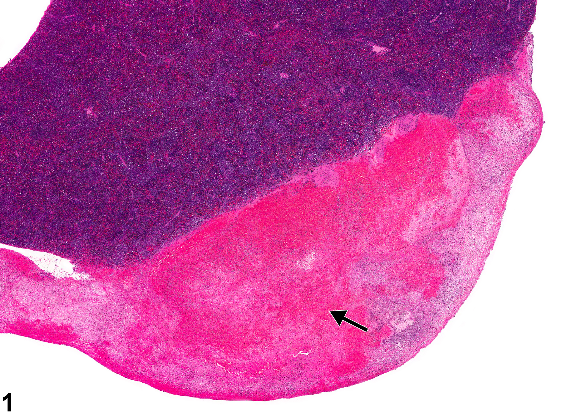
Spleen - Hemorrhage in a female F344/N rat from a chronic study. The splenic parenchyma is markedly expanded by extravasated erythrocytes (arrow).
Figure 2 of 2
next arrow

comment:

Hemorrhage is characterized by the presence of extravasated erythrocytes within the splenic capsule ( Figure 1image opens in a pop-up window , arrow) or parenchyma ( Figure 2image opens in a pop-up window , arrow). Hemorrhage can be focal to diffuse and may occur in a variety of conditions (e.g., splenic infarction), following chemical exposure, secondary to irradiation or traumatic injury, and in association with malignant neoplasms, especially hemangiosarcoma. A splenic hematoma is a localized collection of blood outside of blood vessels and is considered a subset of hemorrhage ( Figure 1image opens in a pop-up window , arrow). Hemorrhage in the spleen should be distinguished from angiectasis, congestion, hemangioma, and hemangiosarcoma. Angiectasis is usually a focal lesion in the spleen and may be associated with an area of fibrosis. Its variably sized blood-filled spaces are lined by endothelium. Congestion can be difficult to differentiate from hemorrhage. Congestion is typically confined to the red pulp sinuses and may or may not have an identifiable cause. Hemangiomas and hemangiosarcomas are neoplasms of the splenic vascular endothelium.

recommendation:

Whenever present in the spleen, hemorrhage should be diagnosed and graded. Splenic hematomas should not be diagnosed separately but diagnosed as hemorrhage. Hemorrhage need not be diagnosed if secondary to splenic neoplasia but should be noted in the pathology narrative.

references:

National Cancer Institute. 1979. NCI-CG-TR-153. Bioassay of o-Toluidine Hydrochloride for Possible Carcinogenicity (CAS No. 636-21-5) in F344/N Rats and B6C3F1 Mice (Feed Studies). NCI, Bethesda, MD.
Abstract: https://ntp.niehs.nih.gov/go/10049

National Toxicology Program. 2010. NTP TR-559. Toxicology and Carcinogenesis of 2,3′,4,4′,5-Pentachlorobiphenyl (PCB 118) (CAS No. 31508-00-6) in Female Harlan Sprague-Dawley Rat (Gavage Studies). NTP, Research Triangle Park, NC.
Abstract: https://ntp.niehs.nih.gov/go/33539

Stefanski SA, Elwell MR, Stromberg PC. 1990. Spleen, lymph nodes, and thymus. In: Pathology of the Fischer Rat: Reference and Atlas (Boorman GA, Eustis SL, Elwell MR, Montgomery CA, MacKenzie WF, eds). Academic Press, San Diego, 369-394.

Ward JM, Mann PC, Morishima H, Frith CH. 1999. Thymus, spleen, and lymph nodes. In: Pathology of the Mouse (Maronpot RR, ed). Cache River Press, Vienna, IL, 333-360.