U.S. flag

An official website of the United States government

Dot gov

The .gov means it's official.
Federal government websites often end in .gov or .mil. Before sharing sensitive information, make sure you're on a federal government site.

Https

The site is secure.
The https:// ensures that you are connecting to the official website and that any information you provide is encrypted and transmitted securely.

Skip to Main Navigation
Skip to Page Content
Skip to Atlas Navigation

Spleen - Hyperplasia, Lymphocyte

Image of hyperplasia, lymphocyte in the spleen from a male B6C3F1/N mouse in a chronic study
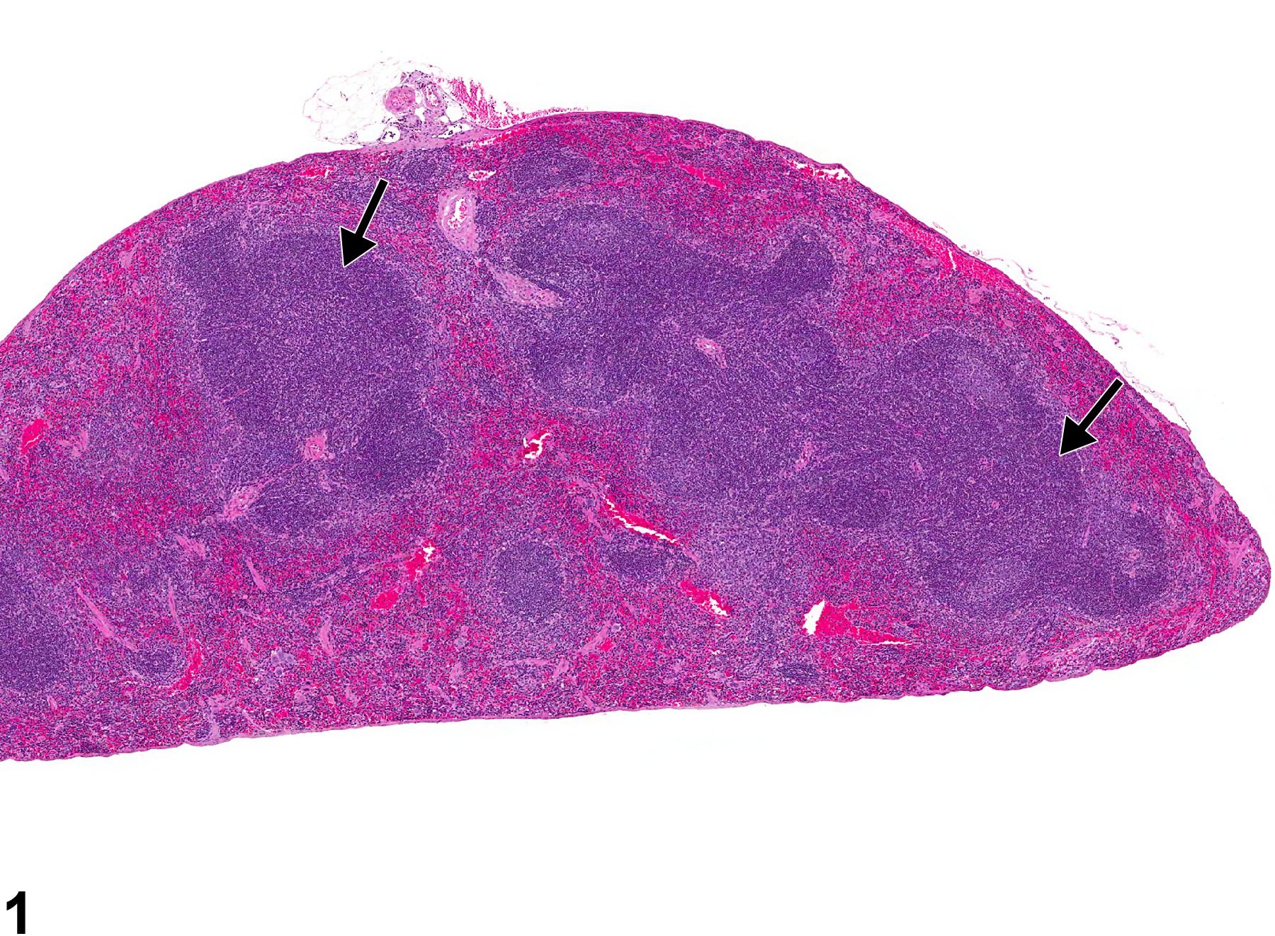
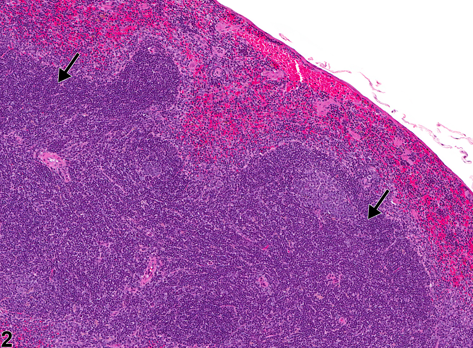
Spleen - Hyperplasia, Lymphocyte in a male B6C3F1/N mouse from a chronic study. The splenic white pulp is expanded by increased numbers of normal lymphocytes (arrows).
Figure 1 of 2
Image of hyperplasia, lymphocyte in the spleen from a male B6C3F1/N mouse in a chronic study
Spleen - Hyperplasia, Lymphocyte in a male B6C3F1/N mouse from a chronic study (higher magnification of Figure 1). Lymphocyte hyperplasia is present in the periarteriolar lymphoid sheaths (arrows).
Figure 2 of 2
next arrow

comment:

Lymphocyte hyperplasia of the spleen is an increase in the number of lymphocytes outside the range of normal compared with concurrent controls. The result can be variable increases in the area and number of white pulp elements (periarteriolar lymphoid sheaths [PALS], follicles, and/or marginal zones) ( Figure 1image opens in a pop-up window and Figure 2image opens in a pop-up window , arrows). This lesion can be focal or generalized. Lymphocyte hyperplasia in the spleen occurs more frequently in mice than in rats, and female mice are more commonly affected than male mice. Marked lymphocyte hyperplasia can be difficult to distinguish from early lymphoma of the spleen; however, the splenic architecture (white pulp and red pulp compartments) should be maintained with lymphocyte hyperplasia ( Figure 1image opens in a pop-up window and Figure 2image opens in a pop-up window ), whereas loss of the splenic architecture would be expected with lymphoma. With generalized lymphocyte hyperplasia, there can be confluency of the PALS regions ( Figure 1image opens in a pop-up window ), so cellular morphology should be evaluated to differentiate from lymphoma. Lymphoma consists of monomorphic (or pleomorphic) neoplastic lymphocytes with increased cellular atypia, whereas lymphocyte hyperplasia consists of increased numbers of normal, mature lymphocytes.

recommendation:

Whenever present, lymphocyte hyperplasia of the spleen should be diagnosed and assigned a severity grade. The number of lymphocytes in the spleen is often variable; therefore, the pathologist must carefully examine and determine the range of normal for a study by comparing with concurrent controls. The plane of section should also be taken into consideration (transverse, longitudinal, tangential, or shallow sections) when evaluating the spleen. Diagnosis should be based upon the pathologist’s best judgment.

references:

Elmore SA. 2006. Enhanced histopathology of the spleen. Toxicol Pathol 34:648-655.
Full Text: https://www.ncbi.nlm.nih.gov/pmc/articles/PMC1828535/

National Toxicology Program. 2011. NTP TR-570. Toxicology and Carcinogenesis Studies of α,β-Thujone (CAS No. 76231-76-0) in F344/N Rats and B6C3F1 Mice (Gavage Studies). NTP, Research Triangle Park, NC.
Abstract: https://ntp.niehs.nih.gov/go/36137

Stefanski SA, Elwell MR, Stromberg PC. 1990. Spleen, lymph nodes, and thymus. In: Pathology of the Fischer Rat: Reference and Atlas (Boorman GA, Eustis SL, Elwell MR, Montgomery CA, MacKenzie WF, eds). Academic Press, San Diego, 369-394.

Suttie AW. 2006. Histopathology of the spleen. Toxicol Pathol 34:466-503.
Full Text: http://tpx.sagepub.com/content/34/5/466.full.pdf

Ward JM, Mann PC, Morishima H, Frith CH. 1999. Thymus, spleen, and lymph nodes. In: Pathology of the Mouse (Maronpot RR, ed). Cache River Press, Vienna, IL, 333-360.

Ward JM, Rehg JE, Morse HC III. 2012. Differentiation of rodent immune and hematopoietic system reactive lesions from neoplasias. Toxicol Pathol 40:425-434.
Abstract: https://www.ncbi.nlm.nih.gov/pubmed/22215512