

Immune System
Spleen - Necrosis
Narrative
Elmore SA. 2006. Enhanced histopathology of the spleen. Toxicol Pathol 34:648-655.
Full Text: https://www.ncbi.nlm.nih.gov/pmc/articles/PMC1828535/National Toxicology Program. 2002. NTP TR-523. Toxicology and Carcinogenesis Studies of Diisopropylcarbodiimide (CAS No. 693-13-0) in F344/N Rats and B6C3F1 Mice (Dermal Studies). NTP, Research Triangle Park, NC.
Abstract: https://ntp.niehs.nih.gov/go/16207Stefanski SA, Elwell MR, Stromberg PC. 1990. Spleen, lymph nodes, and thymus. In: Pathology of the Fischer Rat: Reference and Atlas (Boorman GA, Eustis SL, Elwell MR, Montgomery CA, MacKenzie WF, eds). Academic Press, San Diego, 369-394.
Suttie AW. 2006. Histopathology of the spleen. Toxicol Pathol 34:466-503.
Full Text: http://tpx.sagepub.com/content/34/5/466.full.pdfWard JM, Mann PC, Morishima H, Frith CH. 1999. Thymus, spleen, and lymph nodes. In: Pathology of the Mouse (Maronpot RR, ed). Cache River Press, Vienna, IL, 333-360.
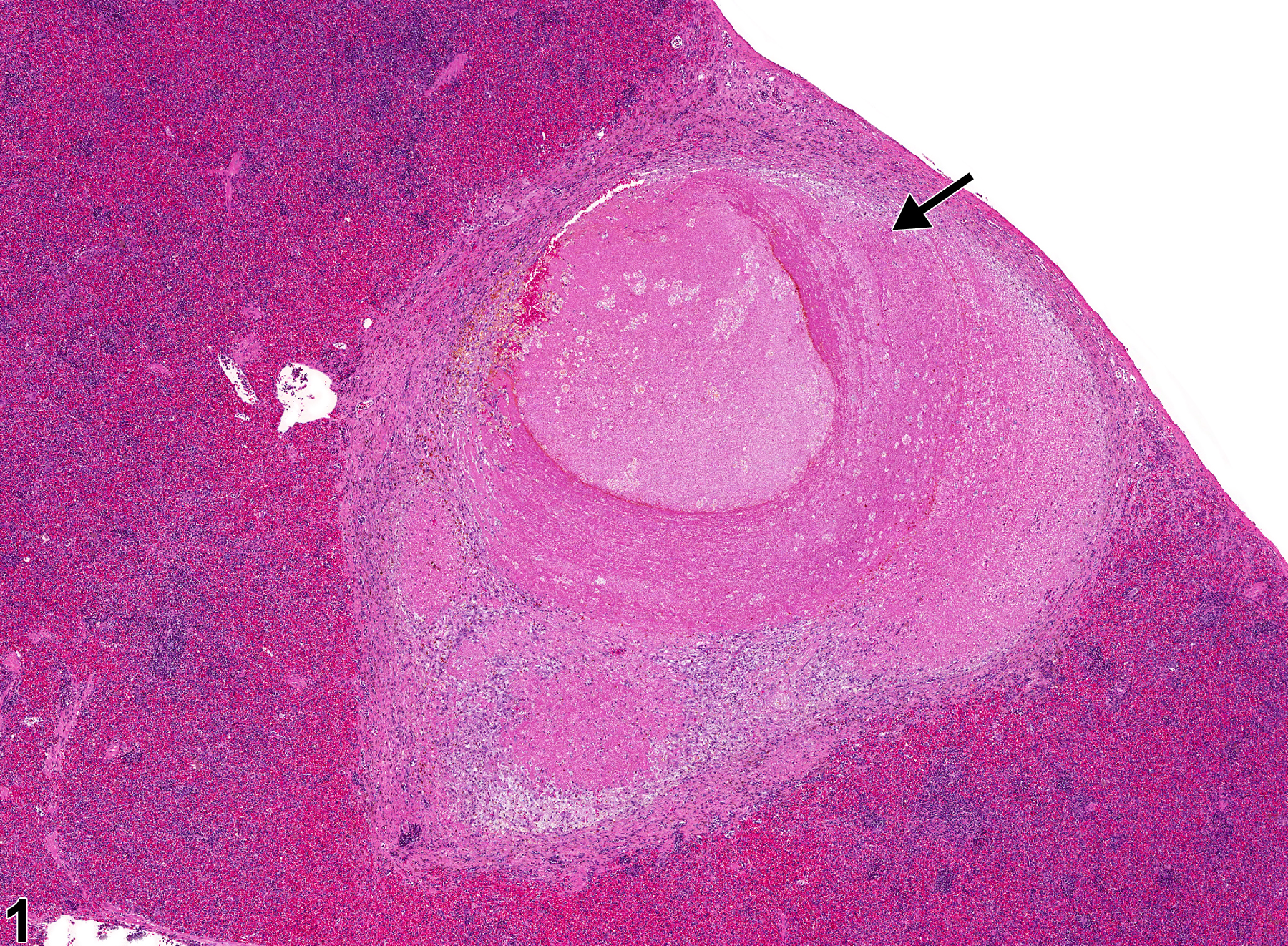
Spleen - Necrosis in a male F344/N rat from a chronic study. A large necrotic focus (arrow) is present with the spleen.
All Images

Spleen - Necrosis in a male F344/N rat from a chronic study. A large necrotic focus (arrow) is present with the spleen.

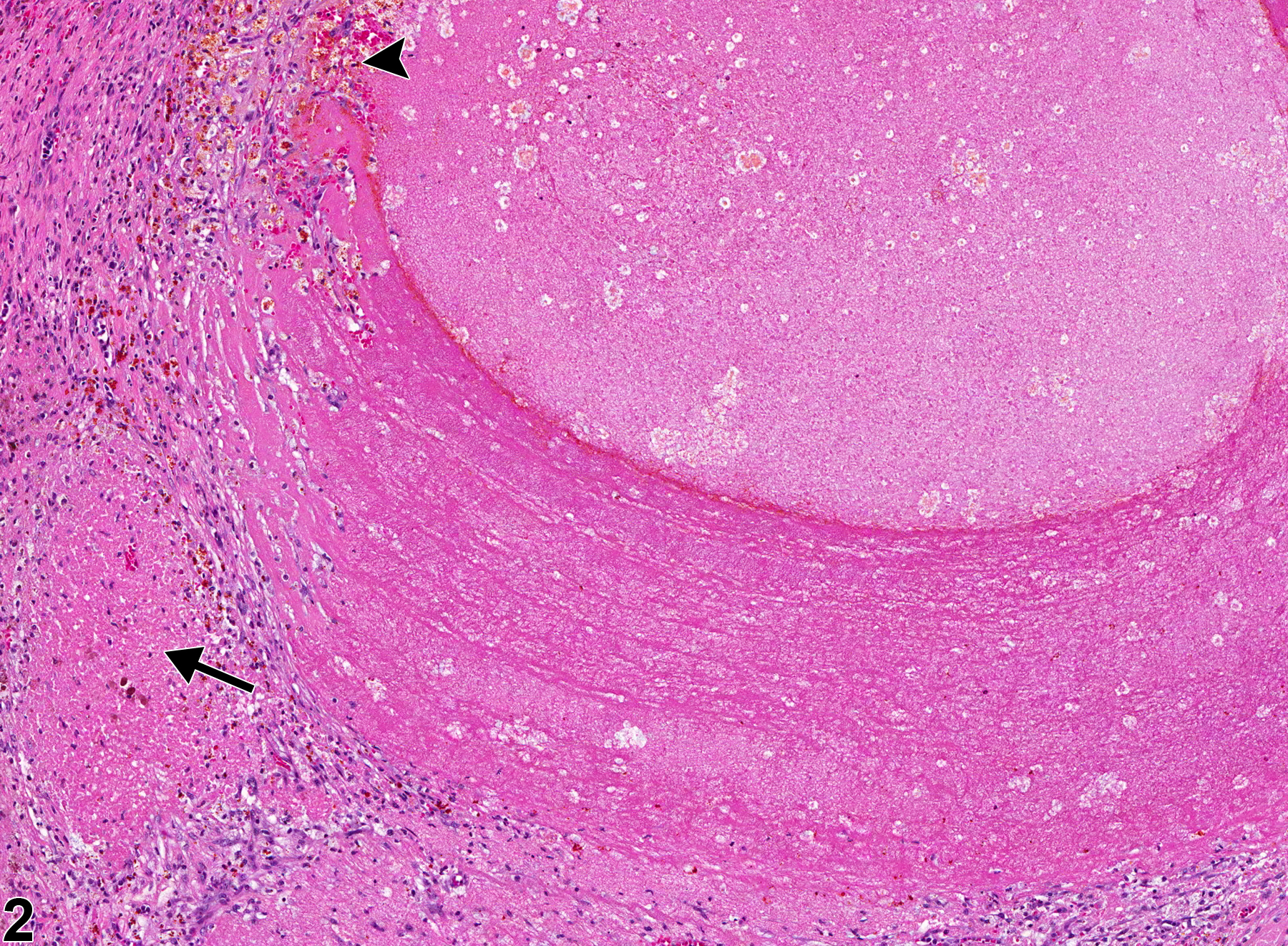
Spleen - Necrosis in a male F344/N rat from a chronic study (higher magnification of Figure 1). Necrosis is bordered by foci of hemorrhage (arrowhead) and fibrous connective tissue (arrow).

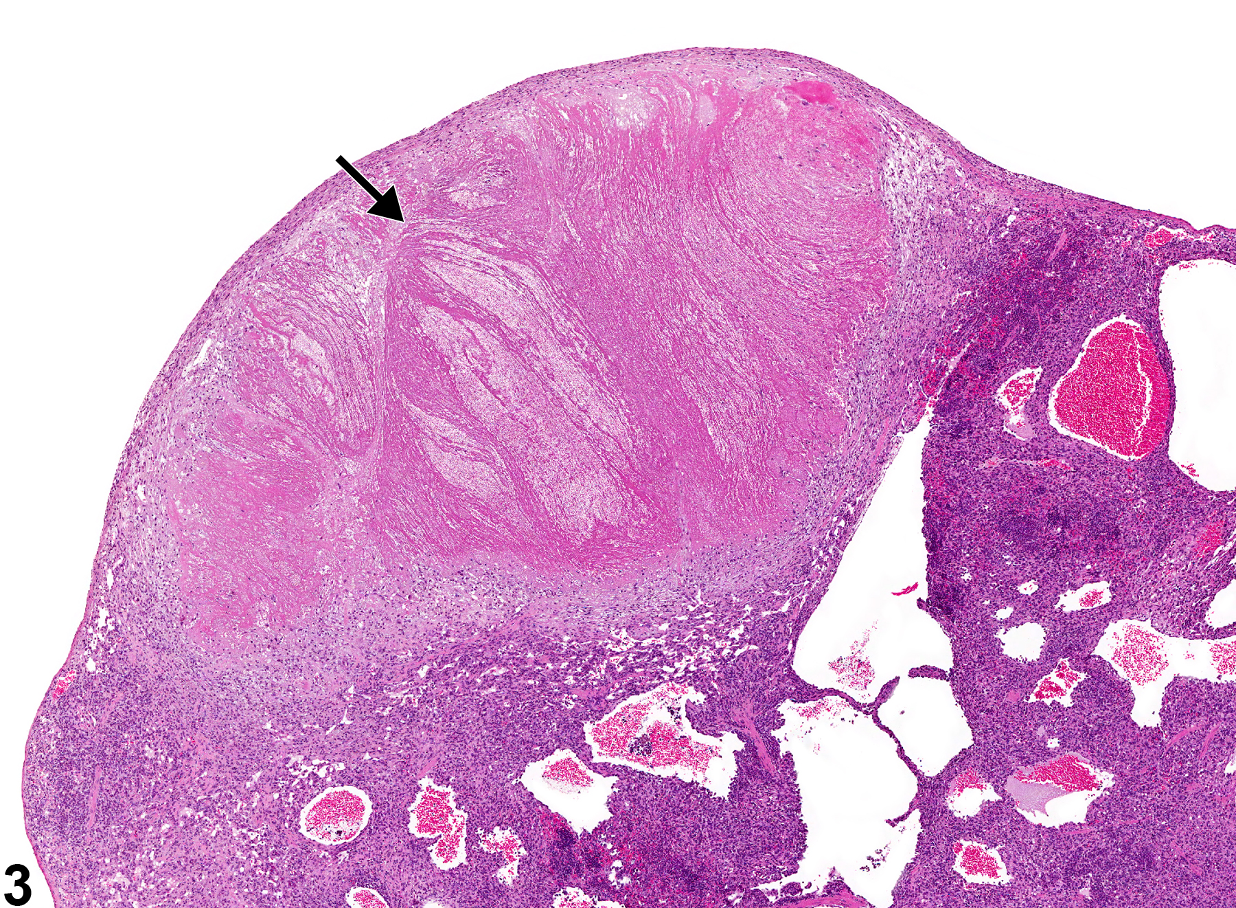
Spleen - Necrosis in a male B6C3F1/N mouse from a chronic study. A large necrotic focus (arrow) is present with the spleen.

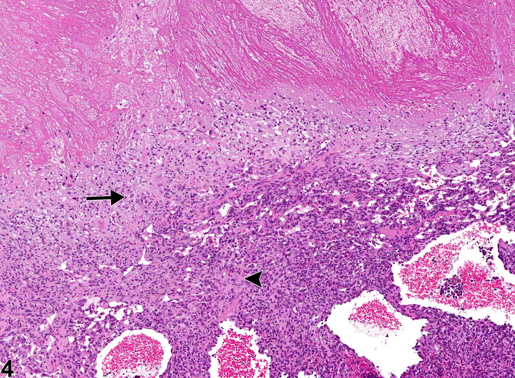
Spleen - Necrosis in a male B6C3F1/N mouse from a chronic study (higher magnification of Figure 3). The necrotic focus is bordered by a mixed inflammatory infiltrate (arrow), fibrosis (arrowhead), and dilated blood vessels.

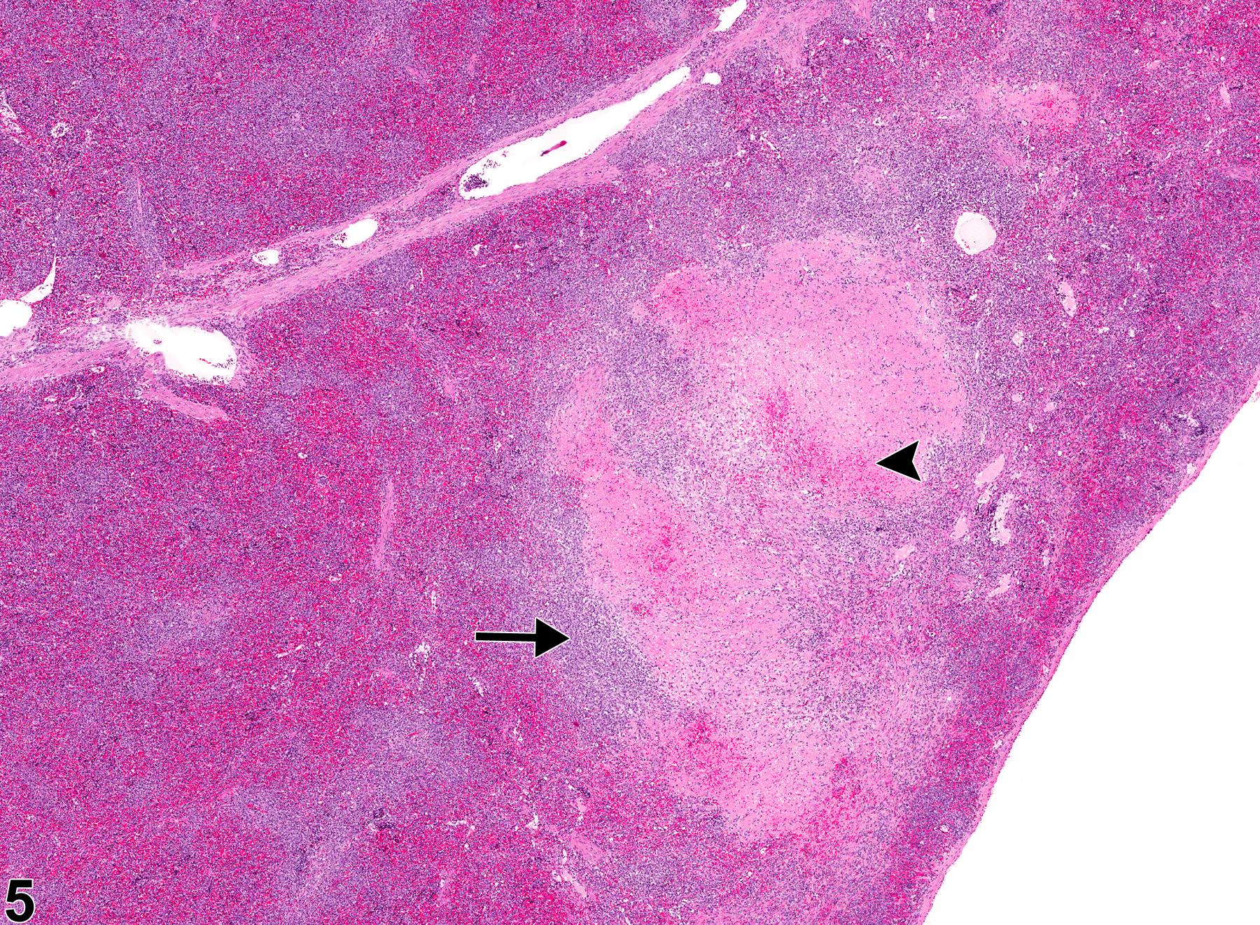
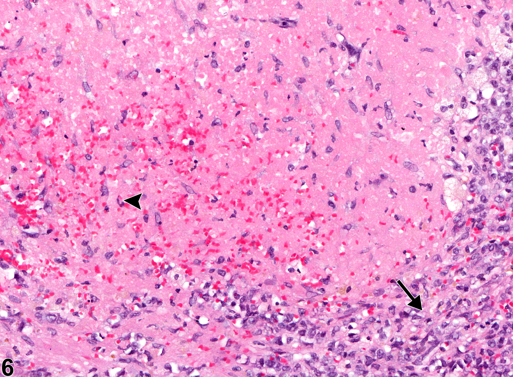
Spleen - Necrosis in a male F344/N rat from a chronic study. A large necrotic focus is present within the splenic red pulp, with focal areas of associated hemorrhage (arrowhead) and bordered by an inflammatory infiltrate (arrow).

Spleen - Necrosis in a male F344/N rat from a chronic study (higher magnification of Figure 5). The necrotic focus consists of necrotic cellular debris, degenerate neutrophils, and hemorrhage (arrowhead) and is bordered by a rim of histiocytes, lymphocytes, and fibroblasts (arrow).