

Immune System
Thymus - Amyloid
Narrative
National Toxicology Program. 2002. NTP TR-507. Toxicology and Carcinogenesis Studies of Vanadium Pentoxide (CAS No. 1314-62-1) in F344/N Rats and B6C3F1 Mice (Inhalation Studies). NTP, Research Triangle Park, NC.
Abstract: https://ntp.niehs.nih.gov/go/14892Peckham JC. 2002. Animal histopathology. In: Handbook of Toxicology, 2nd ed (Derelanko MJ, Hollinger MA, eds). CRC Press, Boca Raton, FL, 685.
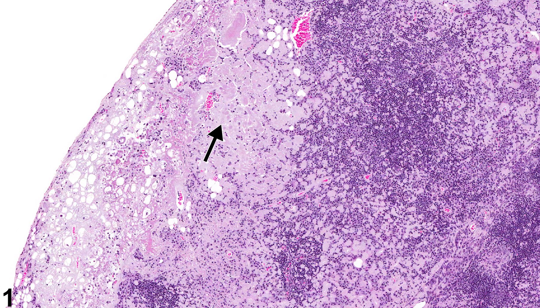
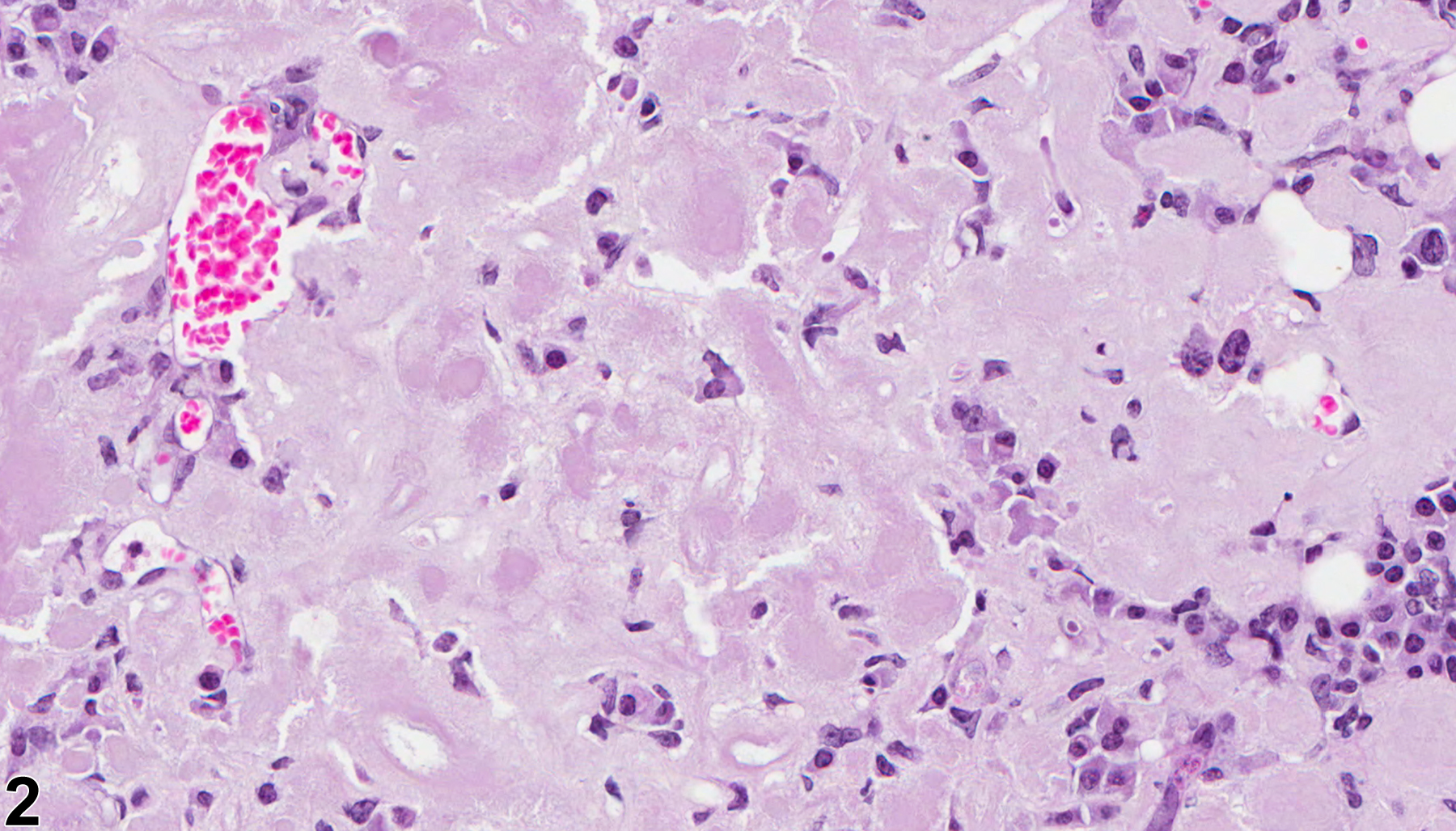
Thymus - Amyloid in a female B6C3F1/N mouse from a chronic study. Normal thymic architecture has been disrupted by deposition of amyloid protein (arrow).
All Images

Thymus - Amyloid in a female B6C3F1/N mouse from a chronic study. Normal thymic architecture has been disrupted by deposition of amyloid protein (arrow).

Thymus - Amyloid in a female B6C3F1/N mouse from a chronic study (higher magnification of Figure 1). Amyloid protein is amorphous, eosinophilic, and extracellular.