U.S. flag

An official website of the United States government

Dot gov

The .gov means it's official.
Federal government websites often end in .gov or .mil. Before sharing sensitive information, make sure you're on a federal government site.

Https

The site is secure.
The https:// ensures that you are connecting to the official website and that any information you provide is encrypted and transmitted securely.

Skip to Main Navigation
Skip to Page Content
Skip to Atlas Navigation

Thymus - Ectopic Tissue

Image of ectopic tissue in the thymus from a male Harlan Sprague-Dawley rat in a subchronic study
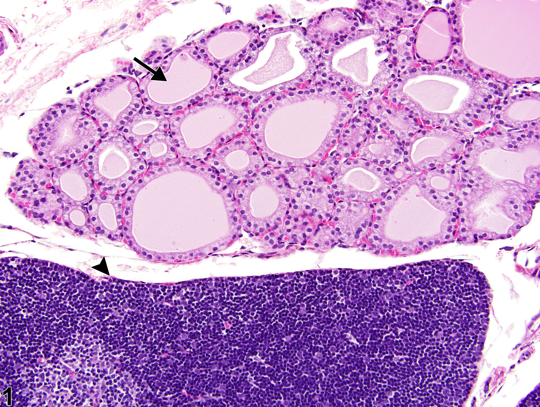
Thymus - Ectopic tissue, Thyroid in a control male Harlan Sprague-Dawley rat from a subchronic study. A structurally normal fragment of thyroid tissue (arrow) is adjacent to the thymus (arrowhead).
Figure 1 of 2
Image of ectopic tissue in the thymus from a female F344/N rat in a chronic study
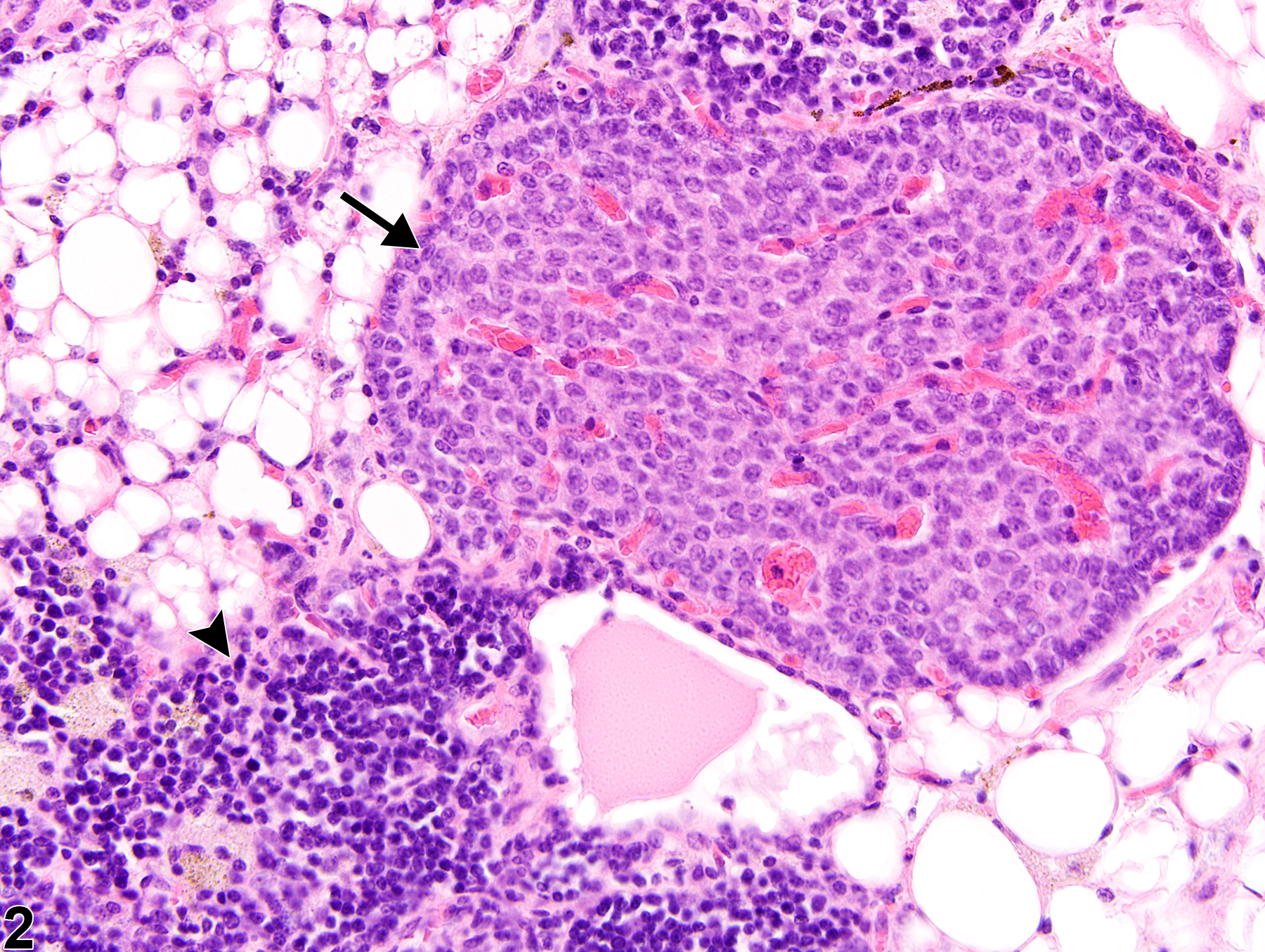
Thymus - Ectopic tissues, Parathyroid in a treated female F344/N rat from a chronic study. Ectopic parathyroid tissue (arrow) extends from the surface of this involuted thymus (arrowhead).
Figure 2 of 2
next arrow

comment:

Ectopic tissue is a fragment of structurally normal tissue (e.g., thyroid, parathyroid) in an abnormal location. This lesion can be congenital or the result of an injury. Ectopic thyroid tissue may occur in and/or adjacent to the thymus of rodents. The thyroid tissue shown in Figure 1image opens in a pop-up window consists of epithelial-lined, colloid-containing follicles separated by a fine fibrovascular stroma. Thyroid C cells may occasionally be found between the follicular epithelium and the basement membrane of the thyroid follicles. Ectopic parathyroid tissue occasionally occurs either adjacent to or embedded in the thymic capsule of rats and/or mice. The parathyroid tissue shown in Figure 2image opens in a pop-up window consists of densely packed, highly folded, branching cords and clusters of polygonal cells separated by a thin fibrovascular stroma. Ectopic parathyroid tissue is an uncommon, incidental finding, although when present it should be distinguished from thymoma or other possible early or small tumors.

recommendation:

Ectopic tissue is typically an incidental finding in or adjacent to the thymus of rodents. When this lesion is present it should diagnosed but not graded. The type of ectopic tissue present (e.g., thyroid, parathyroid) should be included in the diagnosis as a modifier (e.g., Thymus - Ectopic tissue, Thyroid).

references:

Brayton C. 2007. Spontaneous diseases in commonly used mouse strains. In: The Mouse in Biomedical Research: Diseases, 2nd ed (Fox JG, Barthold S, Davisson M, Newcomer CE, Quimby FW, Smith A, eds). Academic Press, Burlington, MA, 676.

National Toxicology Program. 1983. NTP TR-248. Carcinogenesis Studies of 4,4’-Methylenedianiline Dihydrochloride (CAS No. 13552-44-8) in F344/N Rats and B6C3F1 Mice (Drinking Water Studies). NTP, Research Triangle Park, NC.
Abstract: https://ntp.niehs.nih.gov/go/7104

Pearse G. 2006. Histopathology of the thymus. Toxicol Pathol 34:515-547.
Full Text: http://tpx.sagepub.com/content/34/5/515.long

Stefanski SA, Elwell MR, Stromberg PC. 1990. Spleen, lymph nodes, and thymus. In: Pathology of the Fischer Rat: Reference and Atlas (Boorman GA, Eustis SL, Elwell MR, Montgomery CA, MacKenzie WF, eds). Academic Press, San Diego, 369-394.

Ward JM, Mann PC, Morishima H, Frith CH. 1999. Thymus, spleen, and lymph nodes. In: Pathology of the Mouse (Maronpot RR, ed). Cache River Press, Vienna, IL, 333-360.