U.S. flag

An official website of the United States government

Dot gov

The .gov means it's official.
Federal government websites often end in .gov or .mil. Before sharing sensitive information, make sure you're on a federal government site.

Https

The site is secure.
The https:// ensures that you are connecting to the official website and that any information you provide is encrypted and transmitted securely.

Skip to Main Navigation
Skip to Page Content
Skip to Atlas Navigation

Thymus - Involution

Image of involution in the thymus from a female Harlan Sprague-Dawley rat in a chronic study
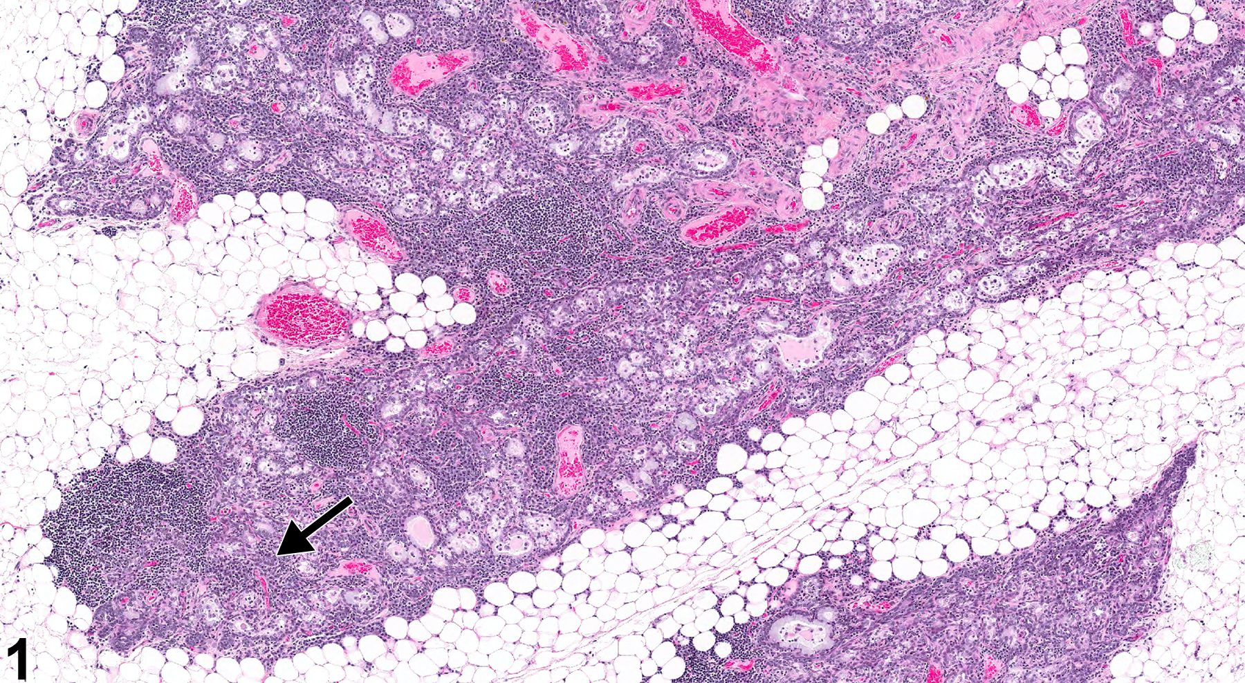
Thymus - Involution in a female Harlan Sprague-Dawley rat from a chronic study. The lymphocyte density is significantly decreased (arrow), and the corticomedullary junction is indistinct. Adipocytes infiltrate the capsule.
Figure 1 of 4
Image of involution in the thymus from a female Harlan Sprague-Dawley rat in a chronic study
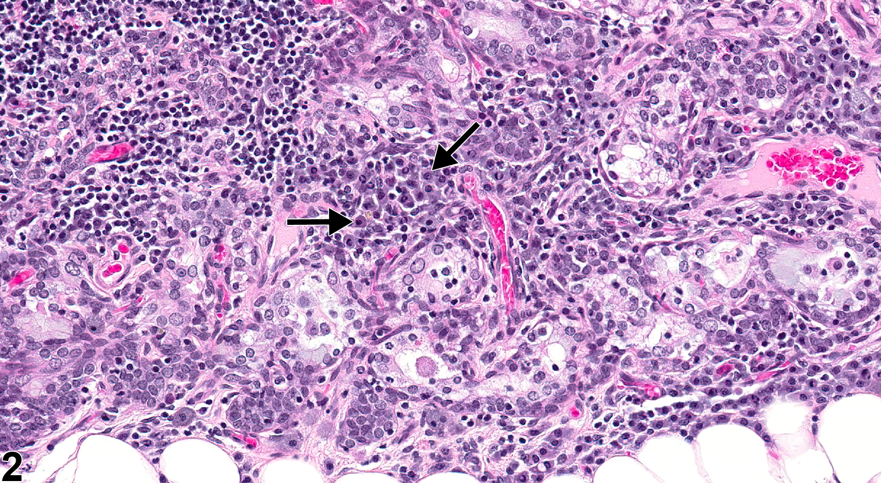
Thymus - Involution in a female Harlan Sprague-Dawley rat from a chronic study (higher magnification of Figure 1). Small aggregates of plasma cells (arrows) are present within the medulla, and medullary epithelial cells are prominent.
Figure 2 of 4
Image of involution in the thymus from a male Wistar Han rat in a chronic study
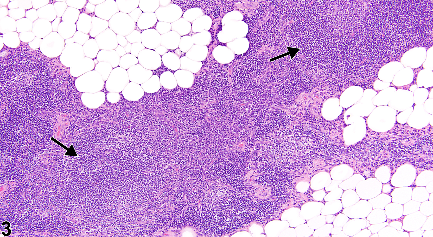
Thymus - Involution in a male Wister Han rat from a chronic study. Adipocytes infiltrate the periphery of the thymus, and the medullary regions are hypercellular (arrows).
Figure 3 of 4
Image of involution in the thymus from a male Wistar Han rat in a chronic study
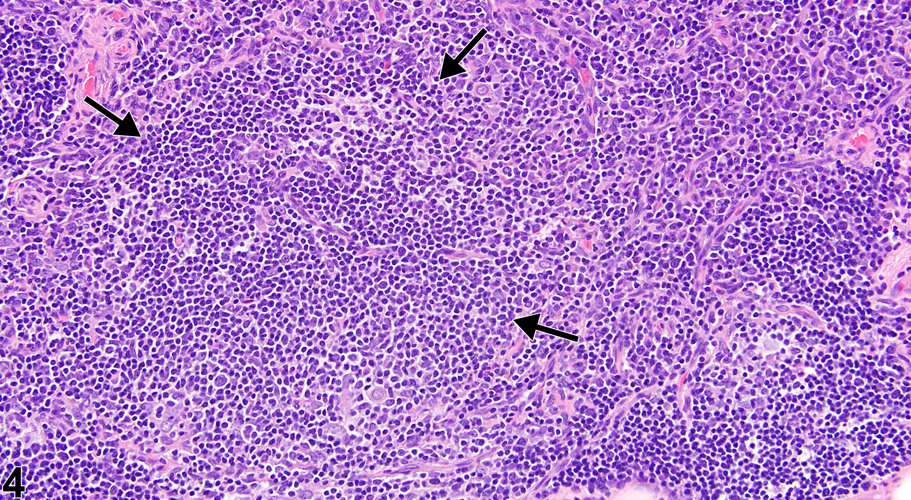
Thymus - Involution in a male Wister Han rat in a chronic study. A focal round area of B-cell hyperplasia is present in the medullary region (arrows).
Figure 4 of 4
next arrow

comment:

Involution of the thymus is a gradual, nonreversible change, likely associated with sex steroid changes that occur during aging. It is a normal physiologic age-related change in rodents and is commonly seen in chronic bioassays. Changes typical of thymus involution include decreased cortical lymphocytes, shrinkage of thymic lobules, thinning and irregularity of the cortex, blurring of the corticomedullary junction, adipocyte infiltration of capsule and septae, increased prominence and/or hyperplasia of medullary epithelial cells, formation of follicular-like B-cell aggregates, cyst formation, and plasma cell hyperplasia. If possible, involution should be distinguished from primary or secondary treatment-related atrophy. Similar to involution, atrophy of the thymus is characterized by reduced thymus size and weight secondary to thymic lymphocyte depletion. However, atrophy in the thymus is unrelated to age, generally caused by toxic insult, and potentially reversible with removal of the inciting agent.

recommendation:

Involution of the thymus is not diagnosed. If possible, normal physiologic age-related involution, which is commonly seen in chronic bioassays, should be distinguished from primary or secondary treatment-related atrophy (see Thymus - Atrophy).

related links:

Thymus - Atrophy

references:

Elmore SA. 2006. Enhanced histopathology of the thymus. Toxicol Pathol 34:656-665.
Full Text: https://www.ncbi.nlm.nih.gov/pmc/articles/PMC1800589/

National Toxicology Program. 2006. NTP TR-525. Toxicology and Carcinogenesis Studies of 2,3,4,7,8-Pentachlorodibenzofuran (PeCDF) (CAS No. 57117-31-4) in Female Harlan Sprague-Dawley Rats (Gavage Studies). NTP, Research Triangle Park, NC.
Abstract: https://ntp.niehs.nih.gov/go/tr525abs

Pearse G. 2006. Histopathology of the thymus. Toxicol Pathol 34:515-547.
Full Text: http://tpx.sagepub.com/content/34/5/515.long