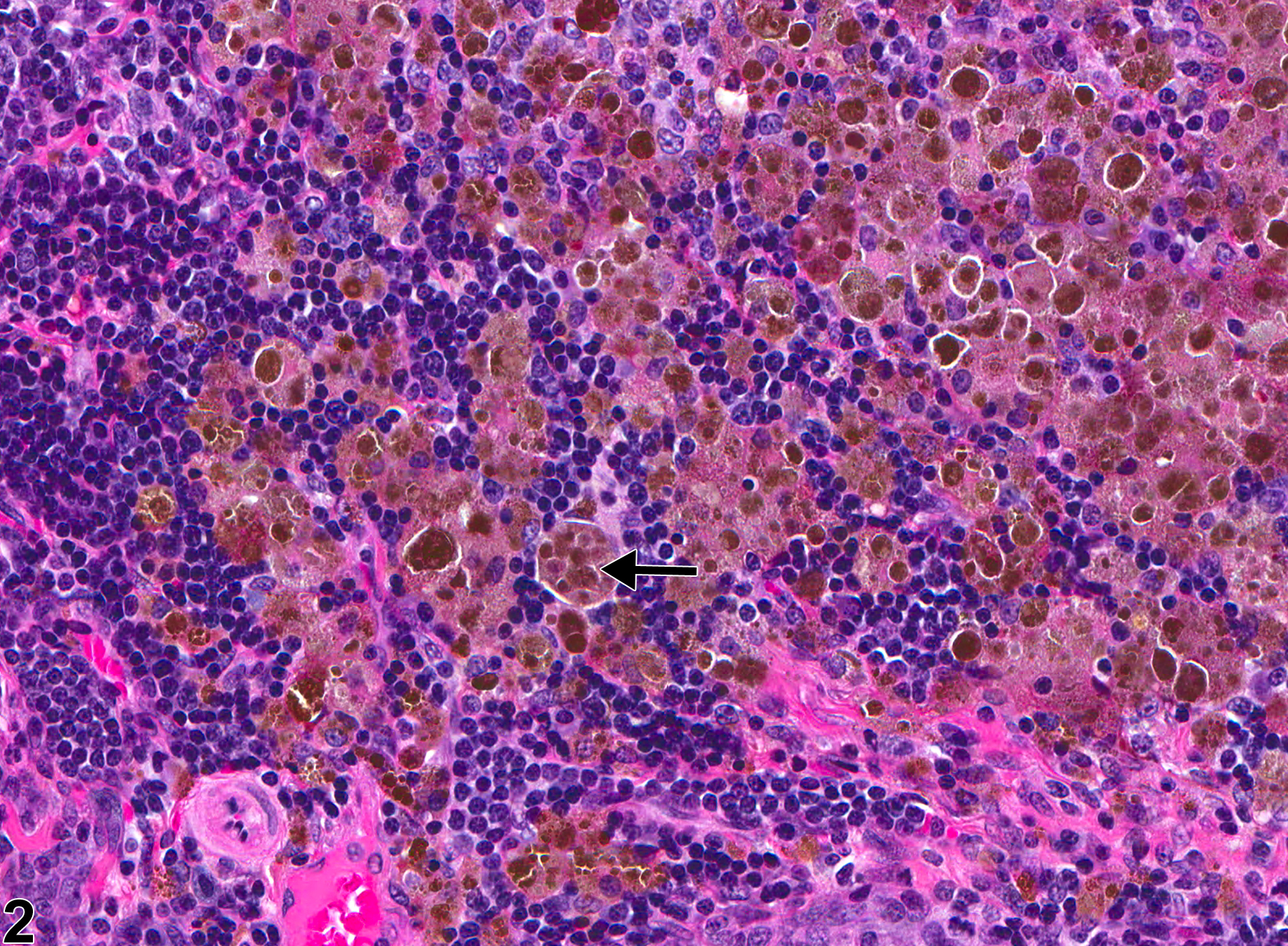
U.S. flag

An official website of the United States government

Dot gov

The .gov means it's official.
Federal government websites often end in .gov or .mil. Before sharing sensitive information, make sure you're on a federal government site.

Https

The site is secure.
The https:// ensures that you are connecting to the official website and that any information you provide is encrypted and transmitted securely.

Skip to Main Navigation
Skip to Page Content
Skip to Atlas Navigation

Thymus - Pigment

Image of pigment in the thymus from a female F344/N rat in a chronic study
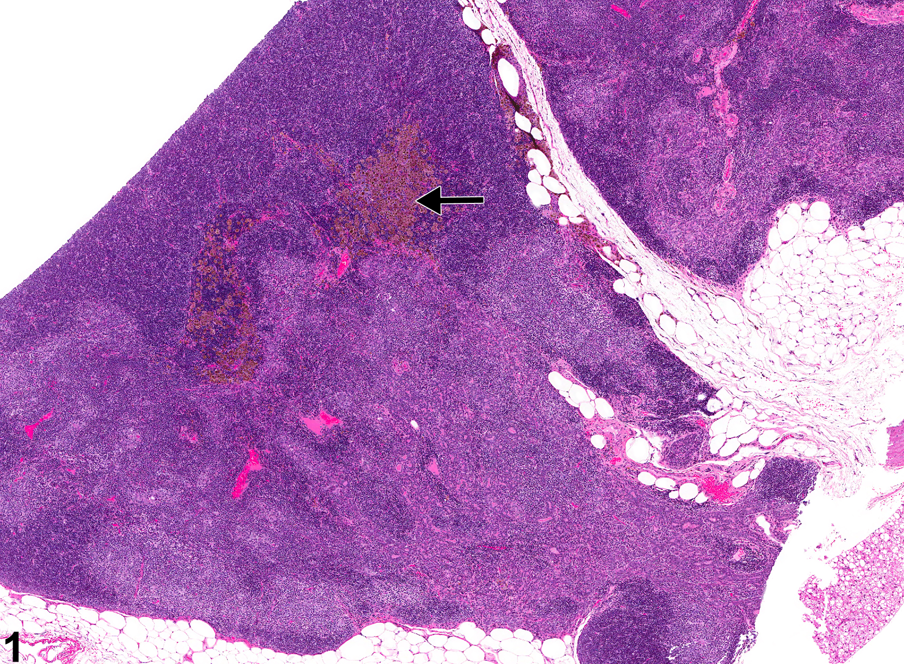
Thymus - Pigment in a female F344/N rat from a chronic study. Brown pigment (likely hemosiderin) (arrow) is present at the thymus corticomedullary junction.
Figure 1 of 2
Image of pigment in the thymus from a female F344/N rat in a chronic study
Thymus - Pigment in a female F344/N rat from a chronic study (higher magnification of Figure 1). Numerous macrophages contain intracytoplasmic dark brown granular pigment (arrow).
Figure 2 of 2
next arrow

comment:

Pigment in the thymus is an uncommon finding and is primarily characterized by the presence of golden to dark brown pigment within macrophages. Pigment may be hemosiderin ( Figure 1image opens in a pop-up window and Figure 2image opens in a pop-up window ) or ceroid/lipofuscin. Hemosiderin is an iron-containing golden-brown granular material, and macrophages containing this pigment are typically observed with sinus erythrocytosis, congestion, or hemorrhage. Lipofuscin is also a yellow-brown, finely granular pigment; however, it is derived mainly from the breakdown products of lipids, such as cell membranes. Ceroid is a variant of lipofuscin that is acid-fast and autofluorescent. Definitive differentiation of these pigments with conventional hematoxylin and eosin (H&E) staining is not possible. Hemosiderin can be identified with iron stains such as Perl's iron and Prussian blue, both of which stain the pigment blue. Ceroid/lipofuscin can be identified with Sudan black B, Schmorl's reaction, Oil red O, carbol lipofuscin stain, periodic acid-Schiff, Ziehl-Neelsen acid-fast stain, autofluorescence, or lysosomal acid phosphatase and esterase stains.

recommendation:

Pigment in the thymus should be diagnosed and given a severity grade. However, not all pigments have to be diagnosed, as some are ubiquitous in aging animals or related to some other disease process and not toxicologically meaningful. The pathologist should use his or her judgment in deciding whether deposits of pigment are toxicologically significant or prominent enough to warrant a separate diagnosis. Definitive pigment identification is often difficult in histologic sections, even with a battery of special stains. Therefore, it is recommended that a diagnosis of "pigment" (as opposed to diagnosing the type of pigment, e.g., hemosiderin or lipofuscin) is most appropriate. The pathology narrative should describe the morphologic features of the pigmentation.

references:

Elmore SA. 2006. Enhanced histopathology of the thymus. Toxicol Pathol 34:656-665.
Full Text: https://www.ncbi.nlm.nih.gov/pmc/articles/PMC1800589/

National Toxicology Program. 2008. NTP TR-541. Toxicology and Carcinogenesis Studies of Formamide (CAS No. 75-12-7) in F344/N Rats and B6C3F1 Mice (Gavage Studies). NTP, Research Triangle Park, NC.
Abstract: https://ntp.niehs.nih.gov/go/29290