U.S. flag

An official website of the United States government

Dot gov

The .gov means it's official.
Federal government websites often end in .gov or .mil. Before sharing sensitive information, make sure you're on a federal government site.

Https

The site is secure.
The https:// ensures that you are connecting to the official website and that any information you provide is encrypted and transmitted securely.

Skip to Main Navigation
Skip to Page Content
Skip to Atlas Navigation

Skin - Hemorrhage

Image of hemorrhage in the skin from a male F344/N rat in a subchronic study
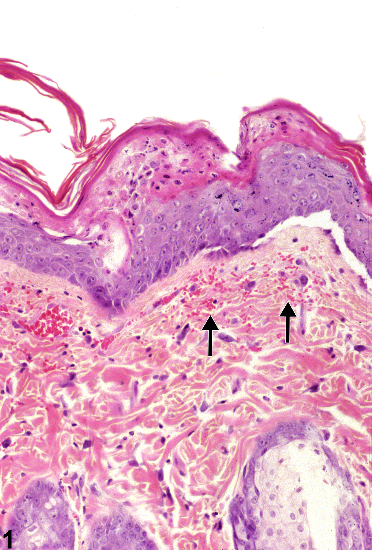
Hemorrhage-extravascular erythrocytes within the dermis (arrows) in a male F344/N rat from a subchronic study.
Figure 1 of 1
next arrow

comment:

Hemorrhage in the skin is characterized by clusters of extravascular erythrocytes ( Figure 1image opens in a pop-up window ). In toxicity and carcinogenicity studies, most spontaneously occurring hemorrhage in the skin is caused by vascular damage due to trauma secondary to fighting (when animals are housed together) or contact with rough edges of equipment. Hemorrhage also occurs as a component of test-article–related skin irritation or damage, often accompanying other lesions such as inflammation, necrosis, and ulceration. Hemorrhage may also be associated with proliferative vascular lesions, such as angiectasis or neoplasia.

recommendation:

In cases where hemorrhage is associated with a proliferative vascular lesion, it should not be recorded. If the hemorrhage is associated with another lesion, such as inflammation or ulceration, it need not be recorded unless the severity of the hemorrhage is excessive or is not commensurate with the severity of the associated lesions. However, it should be described in the pathology narrative. Whenever it is recorded, hemorrhage should be assigned a severity grade.

references:

Elwell MR, Stedman MA, Kovatch RM. 1990. Skin and subcutis. In: Pathology of the Fischer Rat: Reference and Atlas (Boorman GA, Eustis SL, Elwell MR, Montgomery CA, MacKenzie WF, eds). Academic Press, San Diego, 261-277.
Abstract: https://www.ncbi.nlm.nih.gov/nlmcatalog/9002563

Klein-Szanto AJP, Conti CJ. 2002. Skin and oral mucosa. In: Handbook of Toxicologic Pathology, 2nd ed (Haschek WM, Rousseaux CG, Wallig MA, eds). Academic Press, San Diego, 2:85-116.
Abstract: http://www.sciencedirect.com/science/book/9780123302151

Peckham JC, Heider K. 1999. Skin and subcutis. In: Pathology of the Mouse: Reference and Atlas (Maronpot RR, Boorman GA, Gaul BW, eds). Cache River Press, Vienna, IL, 555-612.
Abstract: http://www.cacheriverpress.com/books/pathmouse.htm