

Integumentary System
Skin - Hyperplasia
Narrative
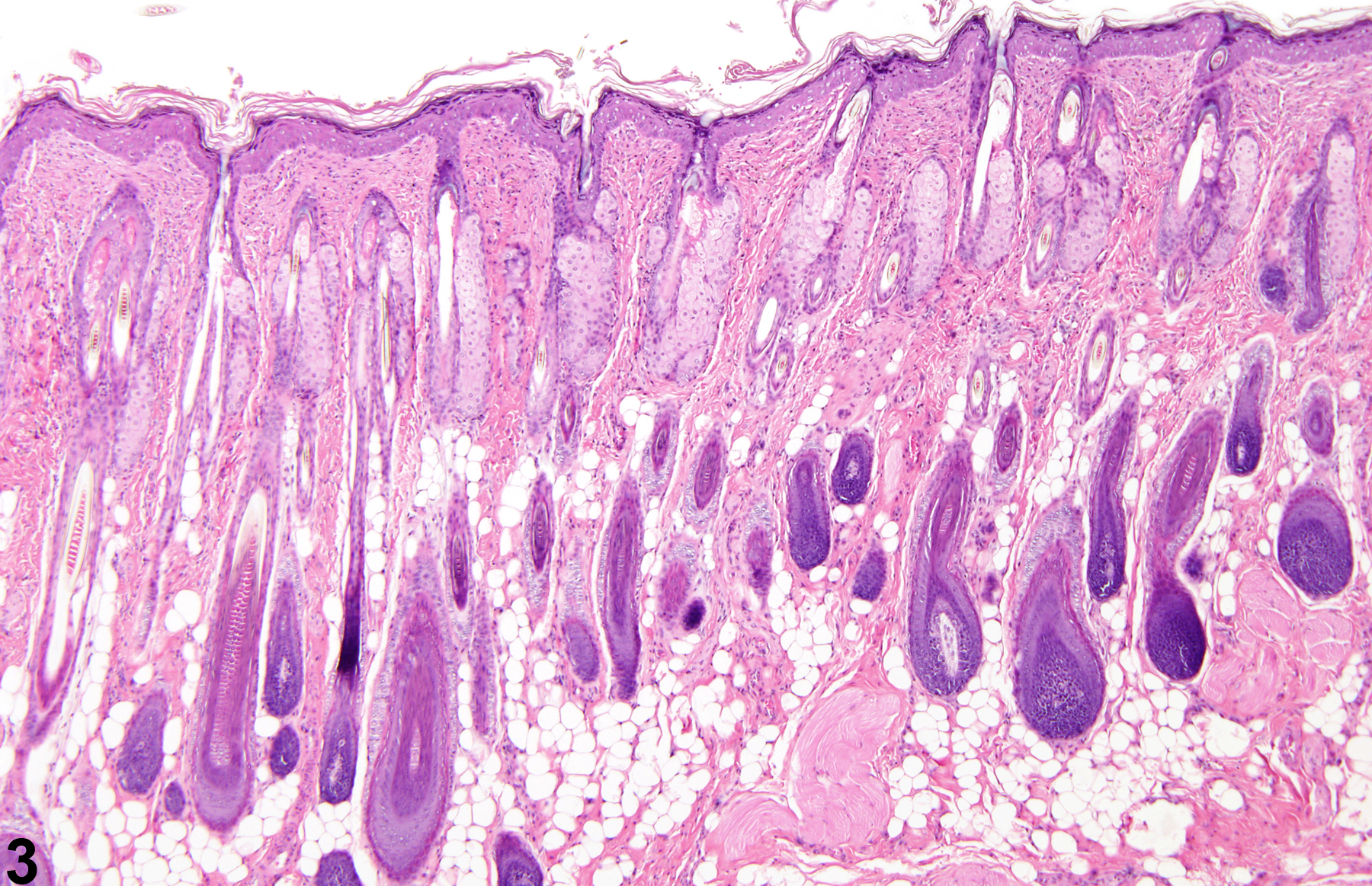
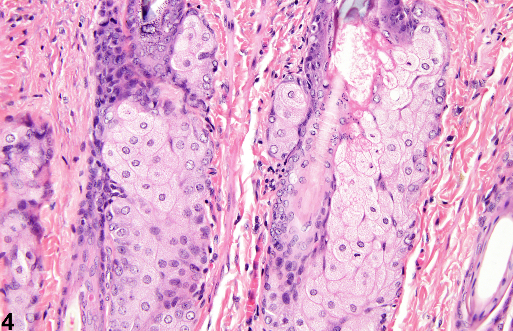
Sebaceous gland hyperplasia is characterized by large sebaceous glands with increased numbers of cells forming numerous lobules around central ducts (Figure 3 and Figure 4). Over the period of a 2-year study, sebaceous hyperplasia has the potential to progress to benign and malignant sebaceous cell neoplasms.
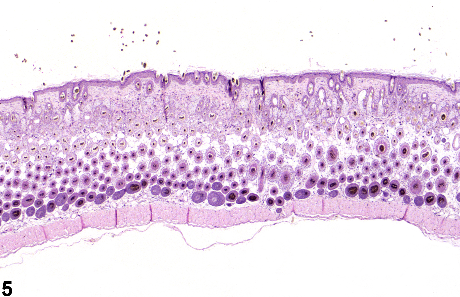
Follicular hyperplasia is characterized by increased numbers of follicular units within the dermis and subcutis (Figure 5). Follicular epithelial hyperplasia is characterized by increased numbers of follicular epithelial cells within the hair follicles; it is typically an extension of epithelial hyperplasia of the epidermis but may be seen in the absence of epidermal hyperplasia.
When present, sebaceous hyperplasia or follicular hyperplasia should be diagnosed and assigned a severity grade. Hyperplasia of the sebaceous cells is often accompanied by hypertrophy. In such cases, hypertrophy need not be diagnosed.
Elwell MR, Stedman MA, Kovatch RM. 1990. Skin and subcutis. In: Pathology of the Fischer Rat: Reference and Atlas (Boorman GA, Eustis SL, Elwell MR, Montgomery CA, MacKenzie WF, eds). Academic Press, San Diego, 261-277.
Abstract: https://www.ncbi.nlm.nih.gov/nlmcatalog/9002563Klein-Szanto AJP, Conti CJ. 2002. Skin and oral mucosa. In: Handbook of Toxicologic Pathology, 2nd ed (Haschek WM, Rousseaux CG, Wallig MA, eds). Academic Press, San Diego, 2:85-116.
Abstract: http://www.sciencedirect.com/science/book/9780123302151Peckham JC, Heider K. 1999. Skin and subcutis. In: Pathology of the Mouse: Reference and Atlas (Maronpot RR, Boorman GA, Gaul BW, eds). Cache River Press, Vienna, IL, 555-612.
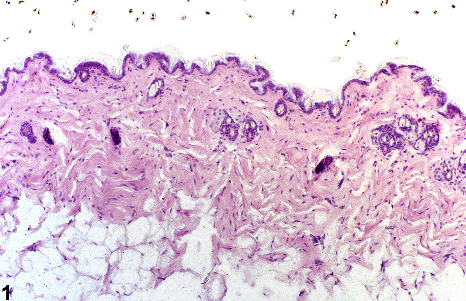
Normal skin in a male B6C3F1 mouse from a 90-day study.
All Images

Normal skin in a male B6C3F1 mouse from a 90-day study.

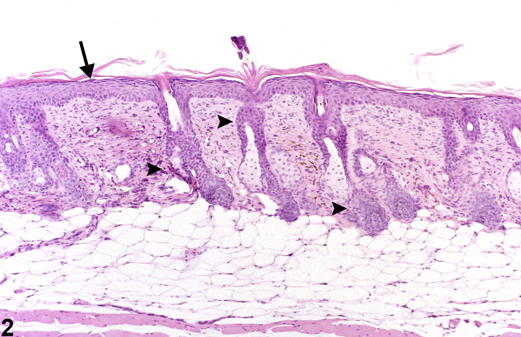
Epithelial hyperplasia-increased numbers of squamous cells in the epidermis (arrow) and follicular epithelium (arrowheads) in a female B6C3F1 mouse from a 90-day study.

Sebaceous gland hyperplasia-increased numbers of sebocytes forming lobules around central ducts in a male F344/N rat from a 90-day study.

Sebaceous gland hyperplasia-increased numbers of sebocytes forming lobules around central ducts in a male F344/N rat from a 90-day study.

Follicular hyperplasia-increased numbers of follicular units within the dermis and subcutis in a female B6C3F1 mouse from a 90-day study.