

Integumentary System
Skin - Pigment
Narrative
Comment:
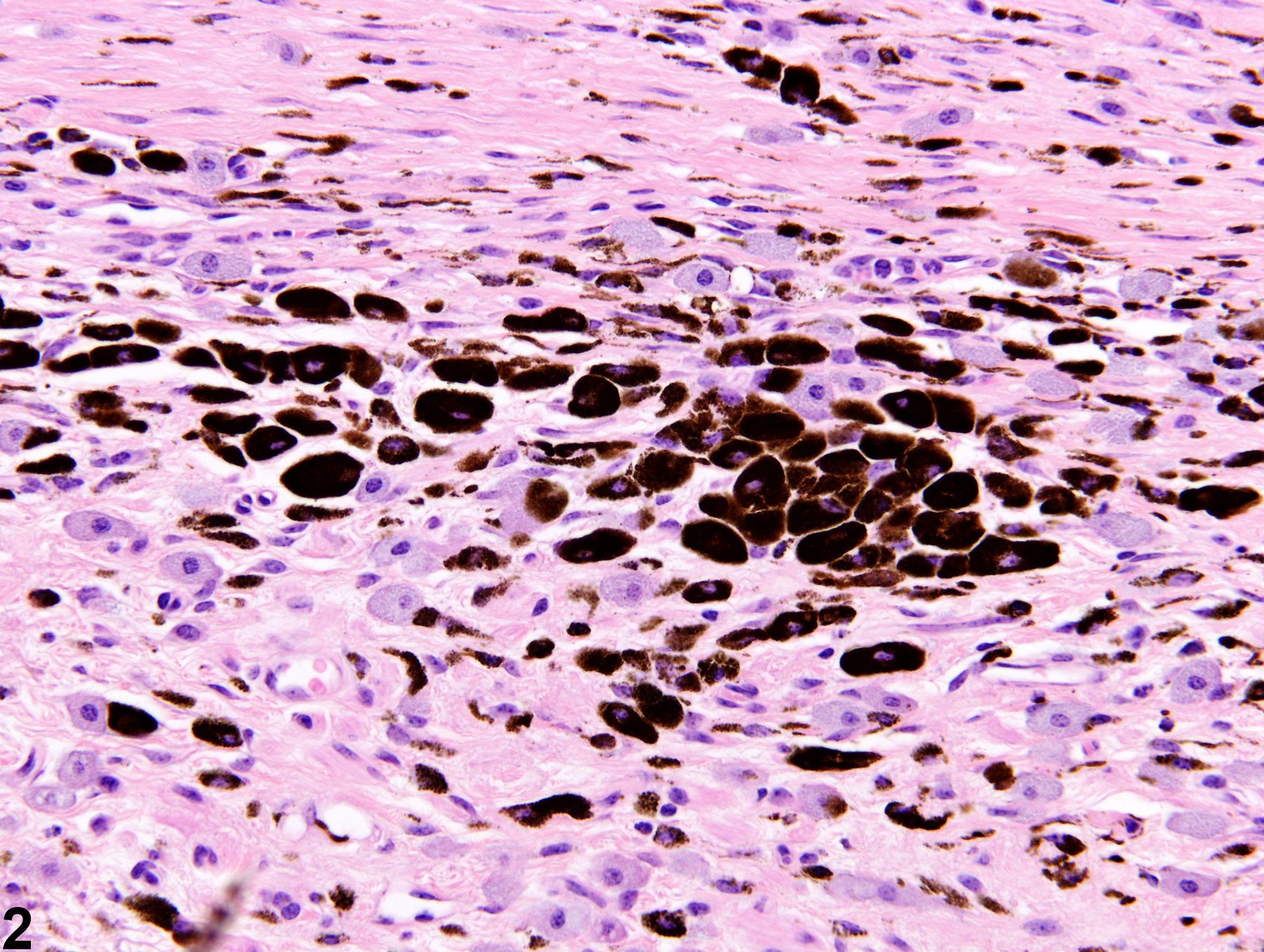
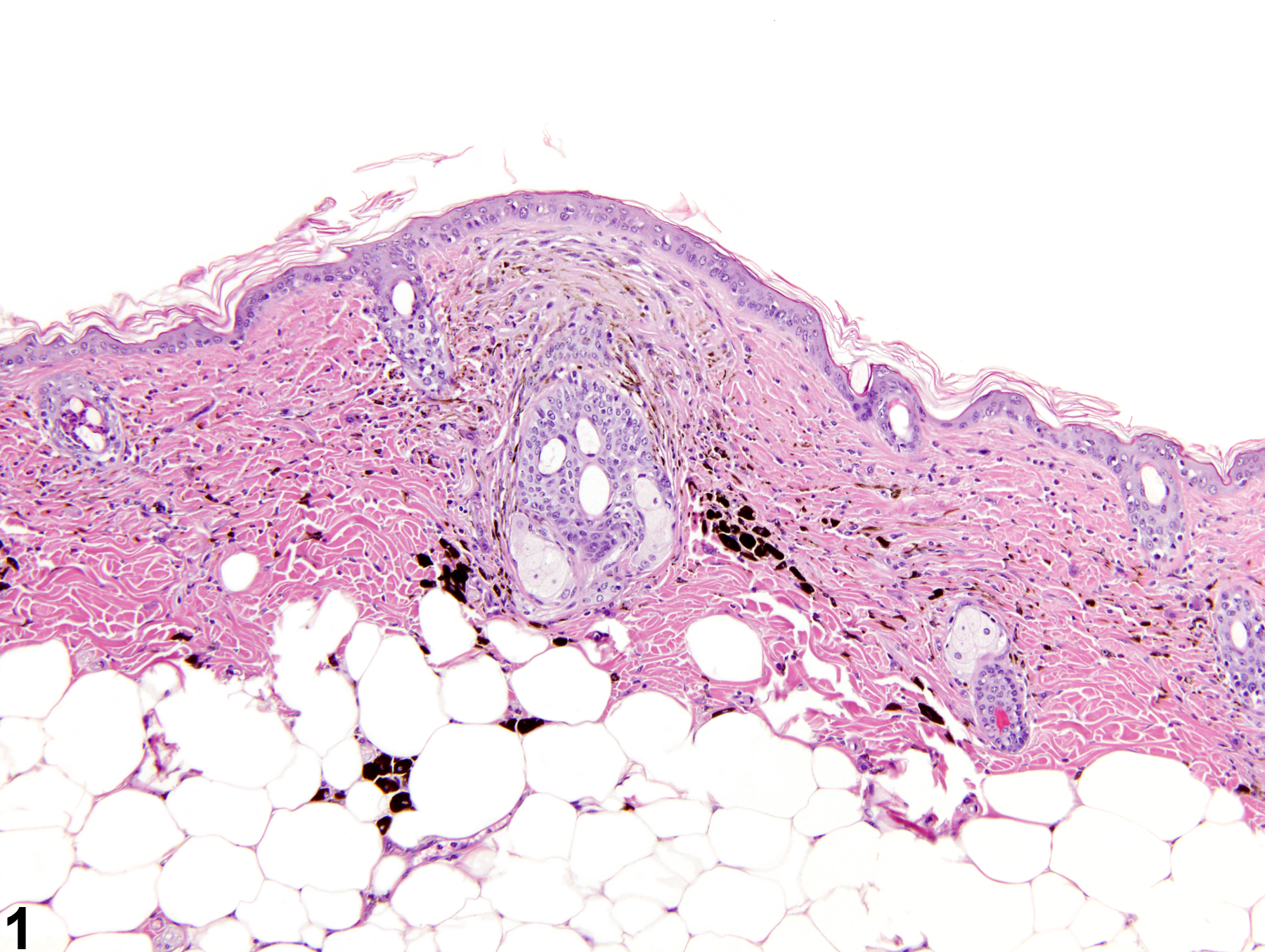
The diagnosis of pigment in the skin is characterized by the accumulation of pigment-laden cells (melanocytes or macrophages) in the dermis or subcutis (Figure 1 and Figure 2). Pigment occurs almost exclusively in dermal studies as part of the response to topical application of a test article. Hence, pigment is typically accompanied by other lesions, such as inflammation (usually chronic or chronic active) or fibrosis.
Recommendations:
When warranted (based on the judgment of the pathologist), pigment should be documented and assigned a severity grade. If it is a minor component of inflammation, it need not be diagnosed separately but should be described in the narrative.
References:
Peckham JC, Heider K. 1999. Skin and subcutis. In: Pathology of the Mouse: Reference and Atlas (Maronpot RR, Boorman GA, Gaul BW, eds). Cache River Press, Vienna, IL, 555-612.
Pigment-accumulation of pigment-laden cells in the dermis in a male B6C3F1 mouse from a chronic study.
All Images

Pigment-accumulation of pigment-laden cells in the dermis in a male B6C3F1 mouse from a chronic study.

Pigment-accumulation of pigment-laden cells in the dermis in a male B6C3F1 mouse from a chronic study.