

Integumentary System
Skin - Pustule
Narrative
Comment:
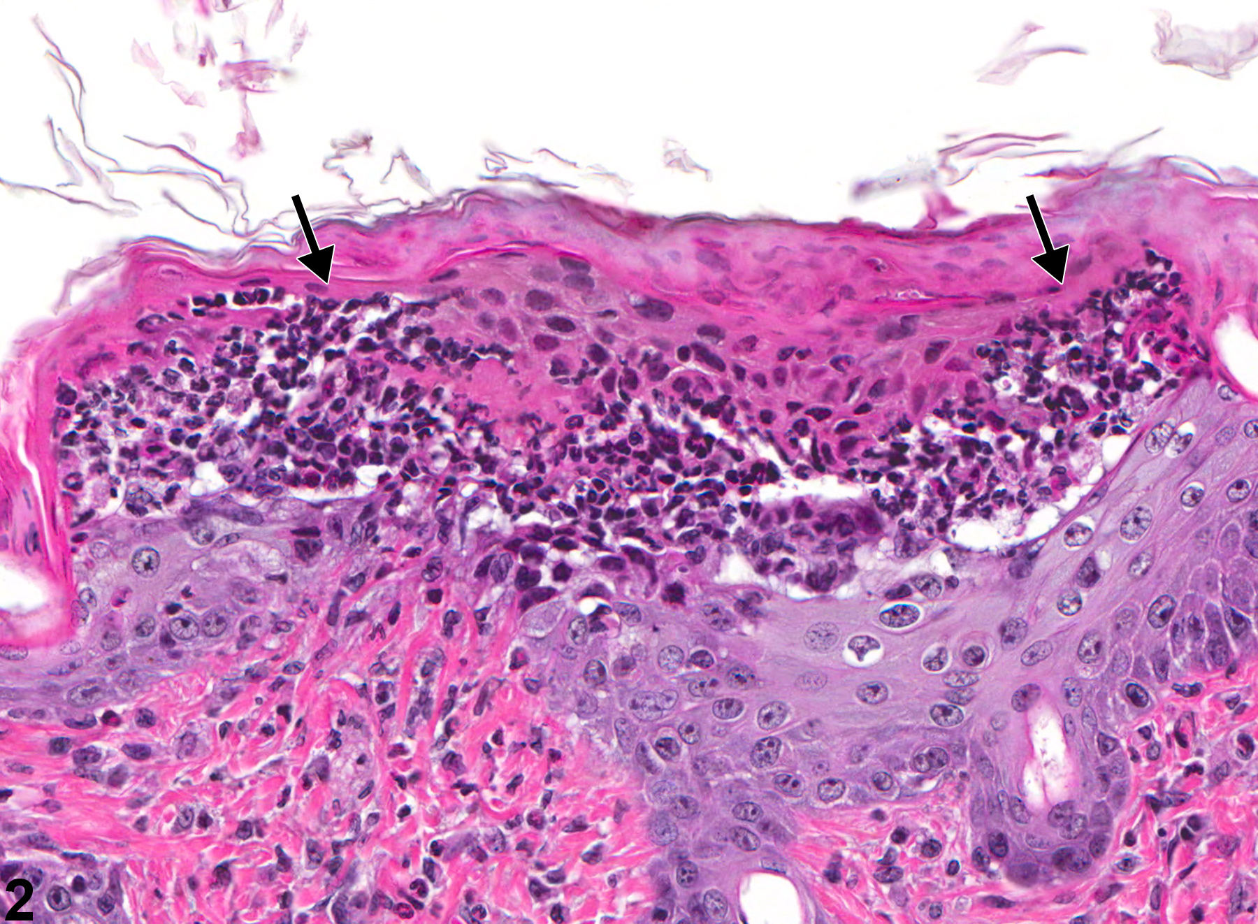
Pustules (Figure 1 and Figure 2) are organized accumulations of neutrophils with or without serum within the stratum corneum. As with vesicles, they may form after exposure to caustic agents. Pustules typically occur in the initial stages of acute inflammation after skin injury but often are not present at the end of a study. Rupture of pustules may result in erosions or ulcers, but since erosions or ulcers do not necessarily arise from pustules, the presence of pustules would provide important information regarding the pathogenesis of the erosions/ulcers.
Recommendations:
Whenever present without apparent underlying erosion or ulceration, pustules should be diagnosed and assigned a severity grade based on size and number. Pustules may be seen with dermal inflammation, epidermal hyperplasia, or hyperkeratosis, all of which should be recorded as separate diagnoses. If pustules are seen concurrently with erosions/ulcers, then both diagnoses should be recorded and graded.
References:
Klein-Szanto AJP, Conti CJ. 2002. Skin and oral mucosa. In: Handbook of Toxicologic Pathology, 2nd ed (Haschek WM, Rousseaux CG, Wallig MA, eds). Academic Press, San Diego, 2:85-116.
Abstract: http://www.sciencedirect.com/science/book/9780123302151National Toxicology Program. 1999. NTP TR 449. Toxicology and Carcinogenesis Studies of Triethanolamine (CAS No. 102-71-6) in F344/N Rats and B6C3F1 Mice (Dermal Studies). NTP, Research Triangle Park, NC.
Abstract: https://ntp.niehs.nih.gov/go/tr449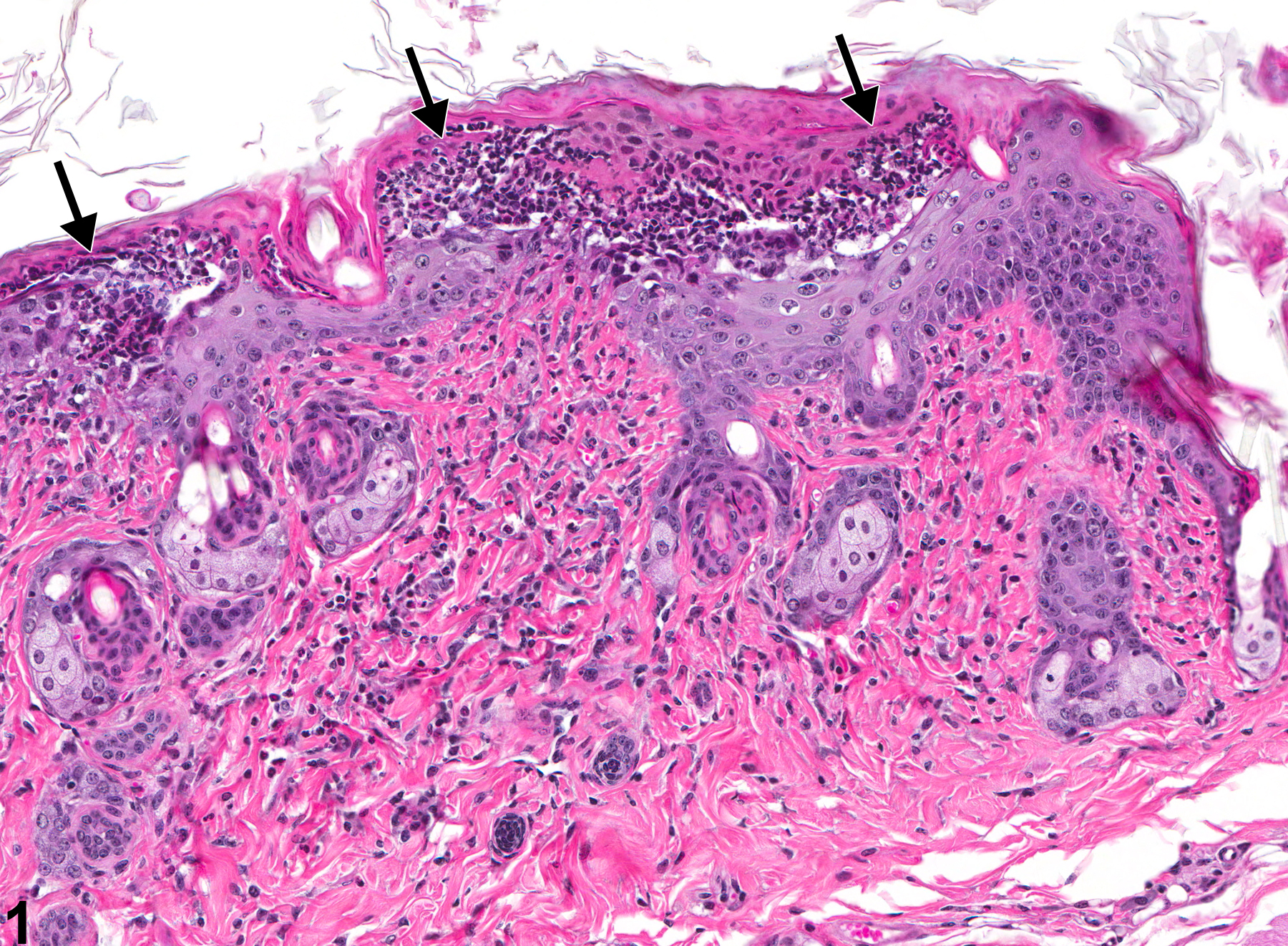
Pustule-accumulations of neutrophils with the stratum corneum (arrows) in a male B6C3F1 mouse from a subchronic study.
All Images

Pustule-accumulations of neutrophils with the stratum corneum (arrows) in a male B6C3F1 mouse from a subchronic study.

Pustule-accumulations of neutrophils with the stratum corneum (arrows) in a male B6C3F1 mouse from a subchronic study.