

Reproductive System, Male
Coagulating Gland - Dilation, Acinar
Narrative
Boorman GA, Elwell MR, Mitsumori K. 1990. Male accessory sex glands, penis, and scrotum. In: Pathology of the Fischer Rat: Reference and Atlas (Boorman GA, Eustis SL, Elwell MR, Montgomery CA, MacKenzie WF, eds). Academic Press, San Diego, 419-428.
Abstract: http://www.ncbi.nlm.nih.gov/nlmcatalog/9002563Gordon LR, Majka JA, Boorman GA. 1996. Spontaneous nonneoplastic and neoplastic lesions and experimentally induced neoplasms of the testes and accessory sex glands. In: Pathobiology of the Aging Mouse, Vol 1 (Mohr U, Dungworth DL, Capen CC, Carlton WW, Sundberg JP, Ward JM, eds). ILSI Press, Washington, DC, 421-441.
Abstract: http://catalog.hathitrust.org/Record/008994685Suwa T, Nyska A, Haseman JK, Mahler JF, Maronpot RR. 2002. Spontaneous lesions in control B6C3F1 mice and recommended sectioning of male accessory sex organs. Toxicol Pathol 30(2):228- 234.
Abstract: http://www.ncbi.nlm.nih.gov/pubmed/11950166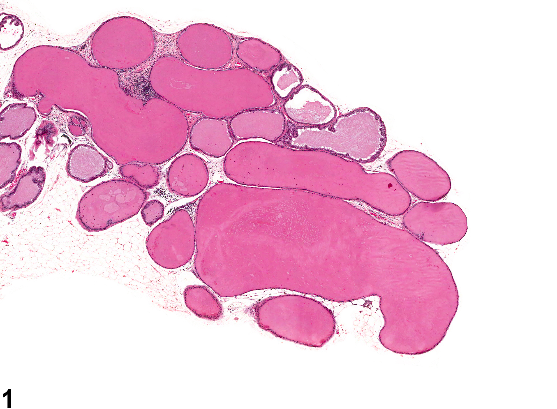
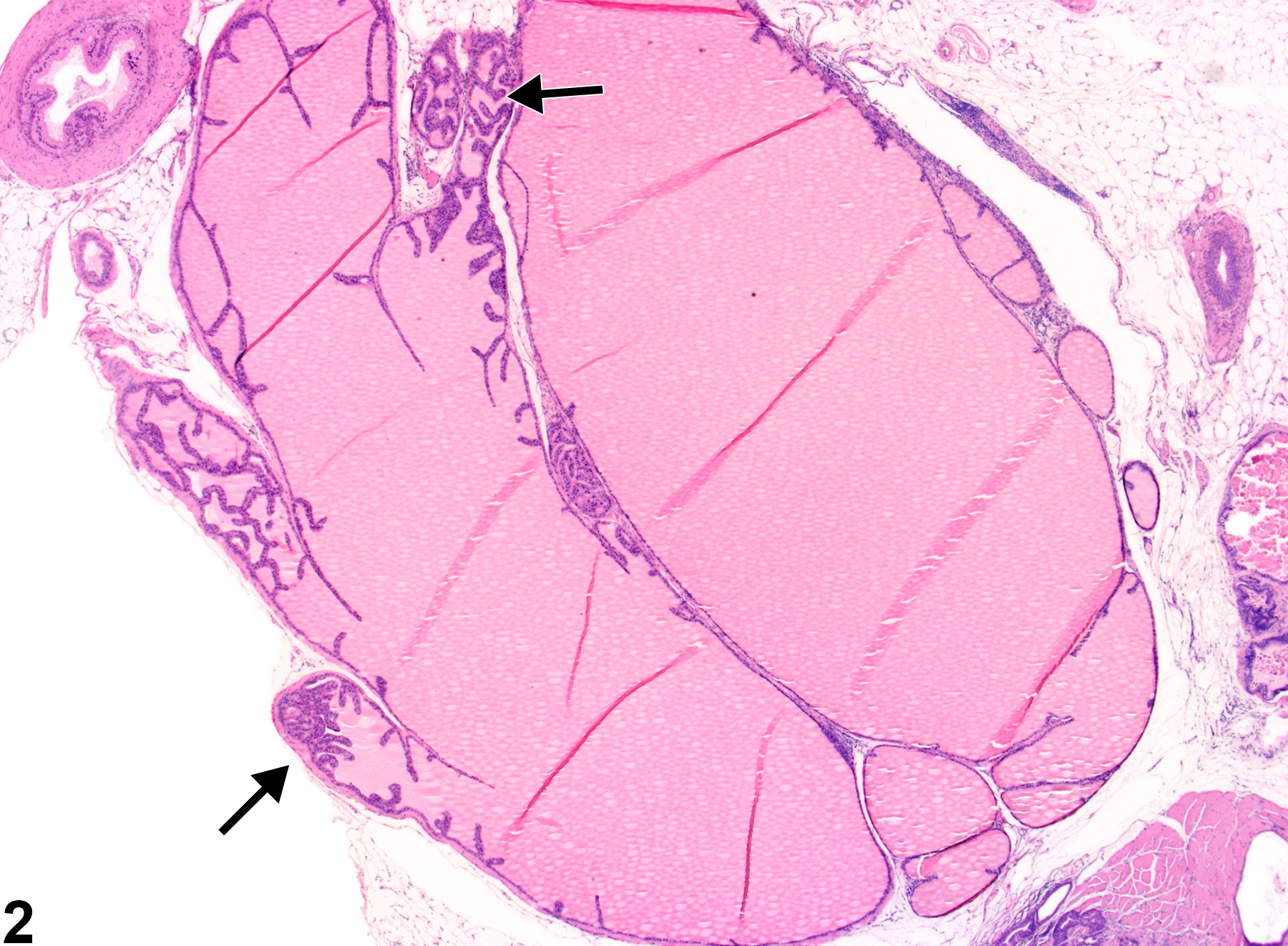
Coagulating Gland - Dilation, Acinar. Acinar dilation of the coagulating gland in a male B6C3F1 mouse from a chronic study.
All Images

Coagulating Gland - Dilation, Acinar. Acinar dilation of the coagulating gland in a male B6C3F1 mouse from a chronic study.

Coagulating Gland - Dilation, Acinar. Collapsed acini with infolding of epithelial structures are present (arrows) in a male B6C3F1 mouse from a chronic study.