

Reproductive System, Male
Coagulating Gland - Hyperplasia
Narrative
Arai Y, Suzuki Y, Nishizuka Y. 1977. Hyperplastic and metaplastic lesions in the reproductive tract of male rats induced by neonatal treatment with diethylstilbestrol. Virchows Arch A Pathol Anat Histol 376:21-28.
Abstract: http://www.ncbi.nlm.nih.gov/pubmed/145081Bosland MC, Tuomari DL, Elwell MR, Shirai T, Ward JM, McConnell RF. 1998. Proliferative lesions of the prostate and other accessory sex glands in male rats, URG-4. In: Guidelines for Toxicologic Pathology. STP/ARP/AFIP, Washington, DC, 1-20.
Pylkkanen L, Santti R, Newbold R, McLachlan JA. 1991. Regional differences in the prostate of the neonatally estrogenized mouse. Prostate 18:117-129.
Abstract: http://www.ncbi.nlm.nih.gov/pubmed/2006118Radovsky A, Mitsumori K, Chapin RE. 1999. Male reproductive tract. In: Pathology of the Mouse: Reference and Atlas (Maronpot RR, Boorman GA, Gaul BW, eds). Cache River Press, Vienna, IL, 381-407.
Strauss L, Makela S, Joshi S, Huhtaniemi I, Santti R. 1998. Genistein exerts estrogen-like effects in male mouse reproductive tract. Mol Cell Endocrinol 144:83-93.
Abstract: http://www.ncbi.nlm.nih.gov/pubmed/9863629Suwa T, Nyska A, Haseman JK, Mahler JF, Maronpot RR. 2002. Spontaneous lesions in control B6C3F1 mice and recommended sectioning of male accessory sex organs. Toxicol Pathol 30(2):228- 234.
Abstract: http://www.ncbi.nlm.nih.gov/pubmed/11950166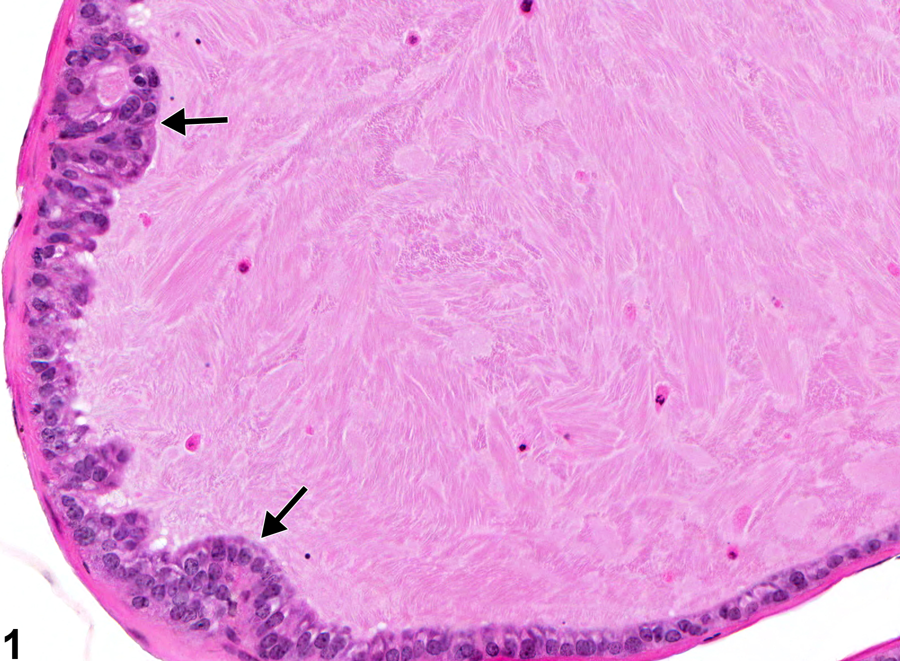
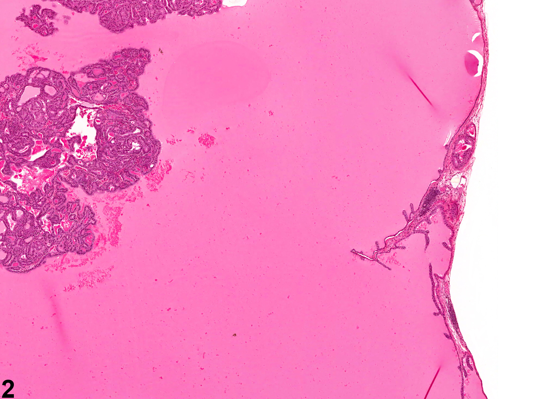
Coagulating Gland - Hyperplasia. Arrows indicate bulging of hyperplastic epithelium into the acinar lumen in a male B6C3F1 mouse from a chronic study.
All Images

Coagulating Gland - Hyperplasia. Arrows indicate bulging of hyperplastic epithelium into the acinar lumen in a male B6C3F1 mouse from a chronic study.

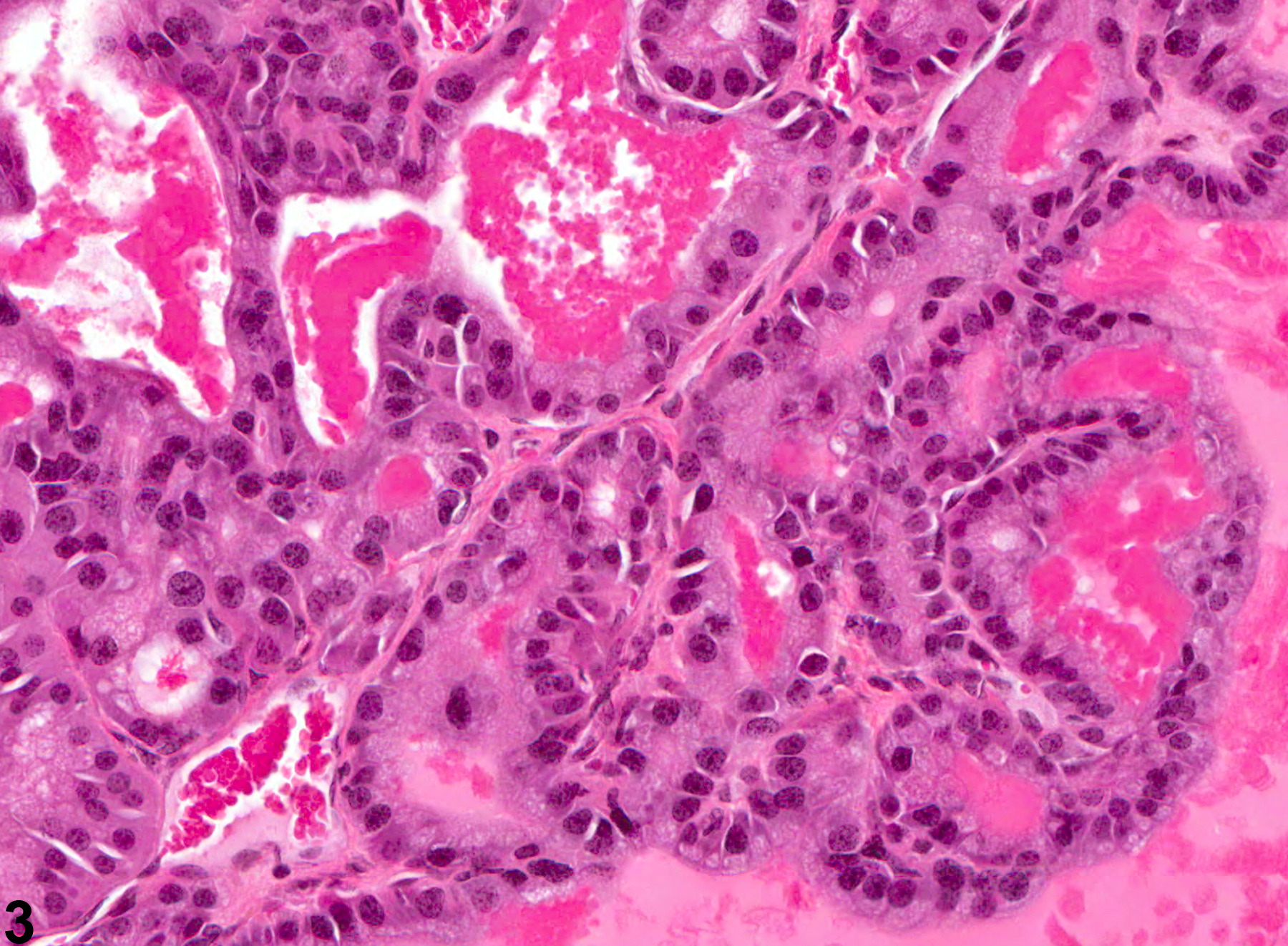
Coagulating Gland - Hyperplasia. Hyperplasia of the coagulating gland in a male B6C3F1 mouse from a chronic study.

Coagulating Gland - Hyperplasia. Higher magnification of Figure 2 showing hyperplasia of the coagulating gland in a male B6C3F1 mouse from a chronic study.