

Reproductive System, Male
Ductus Deferens - Mineralization
Narrative
Comment:
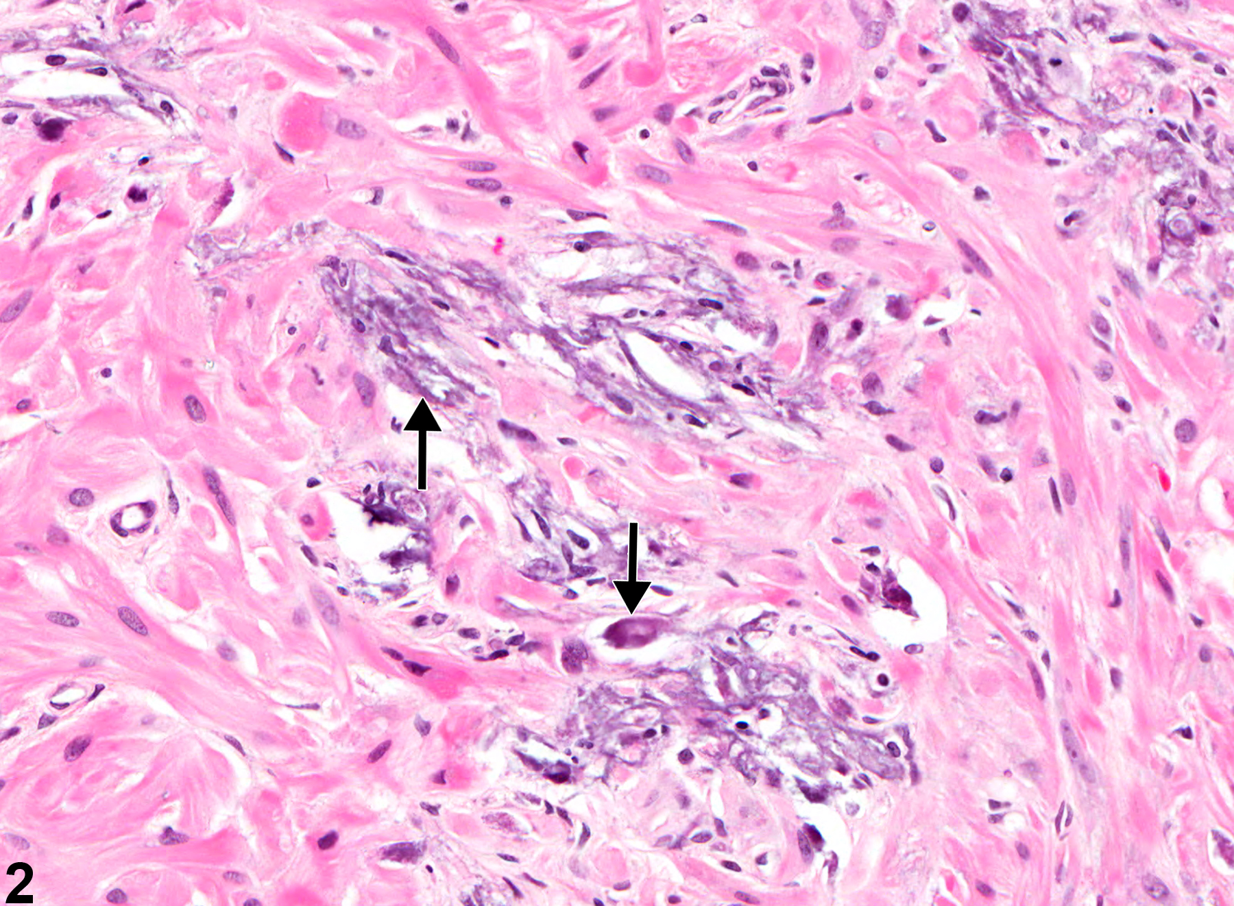
Mineralization is characterized by accumulation of basophilic, fine to coarsely granular to amorphous material (arrows), often without distortion of the histoarchitecture (Figure 1 and Figure 2). Mineralization in the ductus deferens is unusual and is likely an incidental finding not directly associated with chemical treatment. Two types of mineralization may occur: metastatic (calcification of normal tissue associated with high blood levels of calcium) or dystrophic (mineral deposits in abnormal or degenerating tissue not associated with increased blood levels of calcium). When systemic, metastatic mineralization can be present in multiple organs.
Recommendations:
Mineralization should be diagnosed and graded. If it occurs in multiple tissues as a systemic response, it is not necessary to separately diagnose mineralization in the ductus deferens. However, its presence in the ductus deferens may be mentioned in the pathology narrative.
References:
Boorman GA, Elwell MR, Mitsumori K. 1990. Male accessory sex glands, penis, and scrotum. In: Pathology of the Fischer Rat: Reference and Atlas (Boorman GA, Eustis SL, Elwell MR, Montgomery CA, MacKenzie WF, eds). Academic Press, San Diego, 419-428.
Abstract: http://www.ncbi.nlm.nih.gov/nlmcatalog/9002563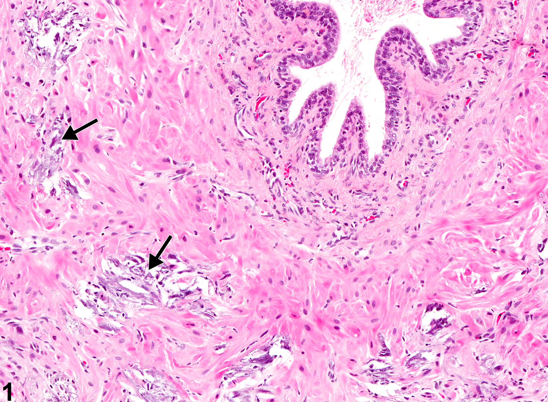
Ductus Deferens - Mineralization. Arrows indicate granular to amorphous mineral deposits in a male B6C3F1 mouse from a chronic study.
All Images

Ductus Deferens - Mineralization. Arrows indicate granular to amorphous mineral deposits in a male B6C3F1 mouse from a chronic study.

Ductus Deferens - Mineralization. Higher magnification of Figure 1. Arrows indicate granular to amorphous mineral deposits in a male B6C3F1 mouse from a chronic study.