U.S. flag

An official website of the United States government

Dot gov

The .gov means it's official.
Federal government websites often end in .gov or .mil. Before sharing sensitive information, make sure you're on a federal government site.

Https

The site is secure.
The https:// ensures that you are connecting to the official website and that any information you provide is encrypted and transmitted securely.

Skip to Main Navigation
Skip to Page Content
Skip to Atlas Navigation

Epididymis, Epithelium - Apoptosis

Image of epithelial apoptosis in the epididymis from a  rat
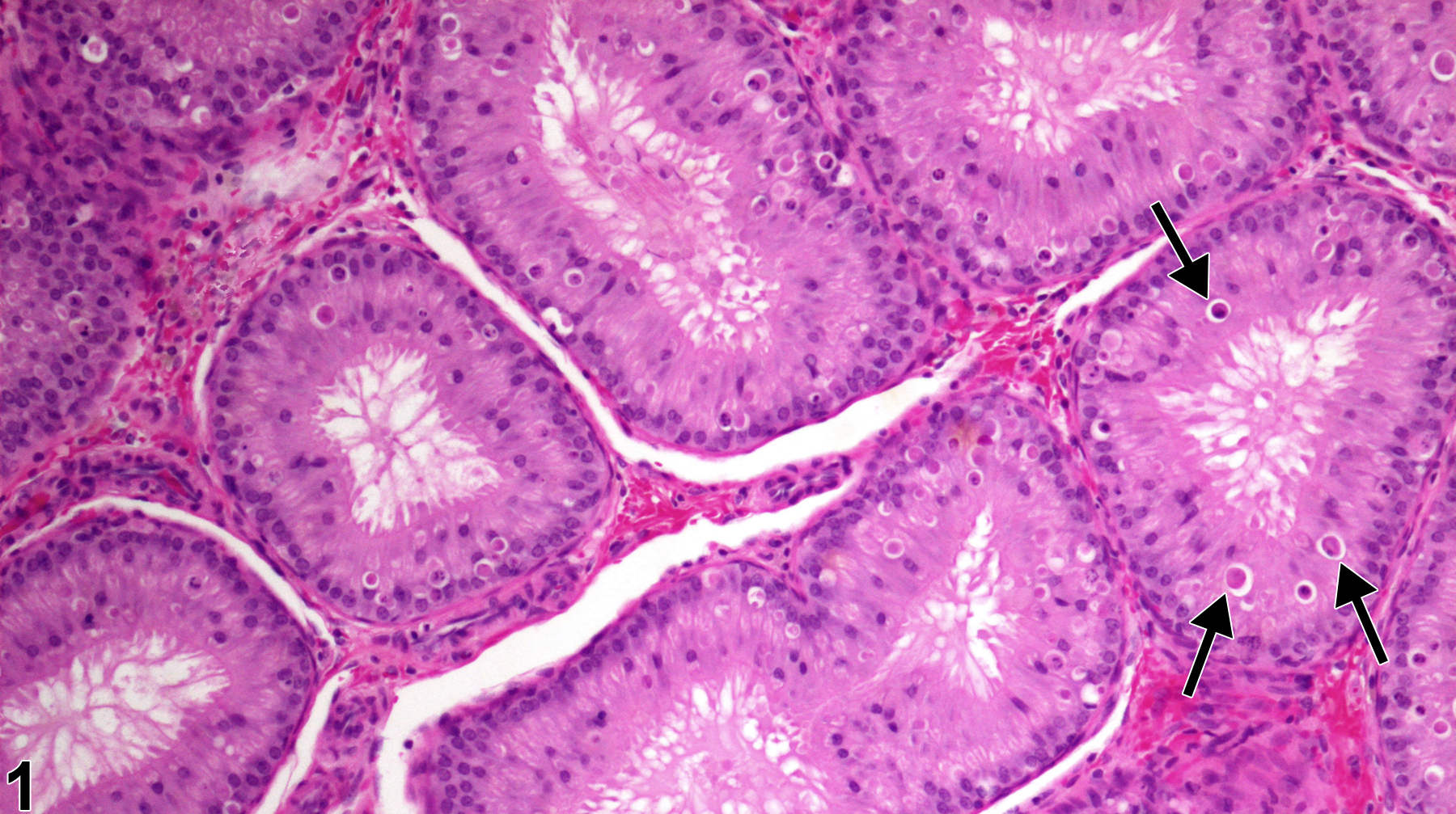
Epididymis, Epithelium - Apoptosis. Numerous apoptotic cells (arrows) are present in the epididymal epithelium of a rat. (Photograph courtesy of D. Creasy.)
Figure 1 of 2
Image of epithelial apoptosis in the epididymis from a  rat
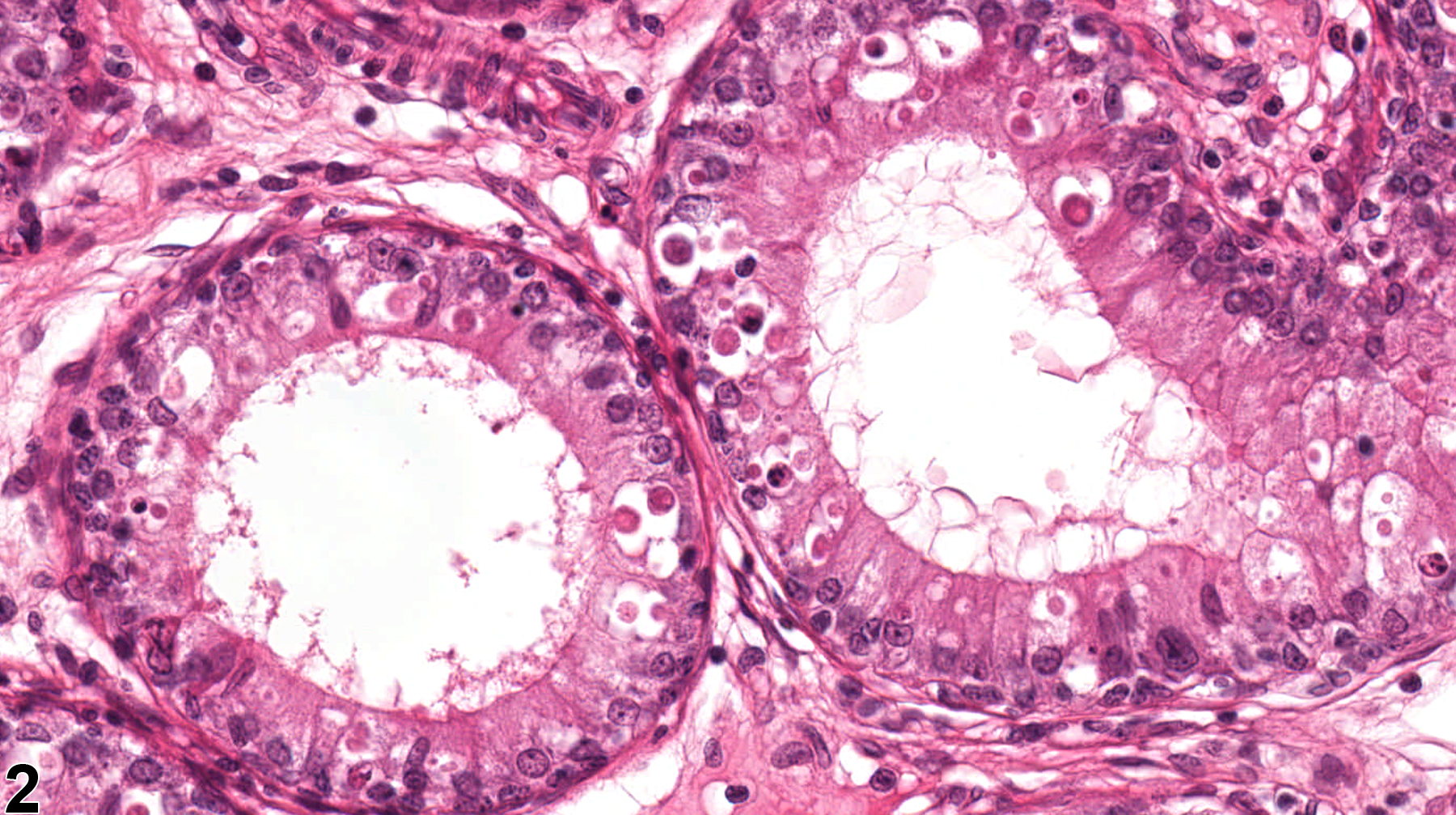
Epididymis, Epithelium - Apoptosis. Numerous apoptotic cells are present within the epithelium lining of the epididymal ducts of a rat. (Photograph courtesy of D. Creasy.)
Figure 2 of 2
next arrow

comment:

Apoptosis (or single cell death) of the epididymal epithelium is most commonly seen as a response to decreased testosterone levels. Normally, there is a low background level of apoptotic cells in the epididymis, but if testosterone levels decrease significantly, the number of apoptotic cells increases, first in the initial segment ( Figure 1image opens in a pop-up window ) and then proceeding distally in a wave-like manner over time through to the cauda. Administration of estrogenic compounds have also been shown to produce this change through interference with testosterone.

recommendation:

Increased levels of apoptotic epithelial cells should be recorded and graded should be and discussed in the pathology narrative if the incidence and/or severity appears to be related to chemical administration. When present in both epididymides, the diagnosis should be clarified as bilateral and the severity score based on the more severely affected epididymis. Correlation with disturbances in other male reproductive organs is recommended to aid interpretation.

references:

Creasy D, Bube A, de Rijk E, Kandori H, Kuwahara M, Masson R, Nolte T, Reams R, Regan K, Rehm S, Rogerson P, Whitney K. 2012. Proliferative and nonproliferative lesions of the rat and mouse male reproductive system. Toxicol Pathol 40:40S-121S.
Abstract: http://www.ncbi.nlm.nih.gov/pubmed/22949412

Ezer N, Robaire B. 2002. Androgenic regulation of the structure and functions of the epididymis. In: The Epididymis: From Molecules to Clinical (Robaire B, Hinton BT, eds). New York, Kluwer/Plenum, 297-316.
Abstract: http://www.springer.com/medicine/urology/book/978-0-306-46684-7

Fan X, Robaire B. 1998. Orchidectomy induces a wave of apoptotic cell death in the epididymis. Endocrinology 139(4):2128-2136.
Abstract: http://www.ncbi.nlm.nih.gov/pubmed/9529002

Robaire B, Fan X. 1998. Regulation of apoptotic cell death in the rat epididymis. J Reprod Fertil Suppl 53:211-214.
Abstract: http://www.ncbi.nlm.nih.gov/pubmed/10645279