U.S. flag

An official website of the United States government

Dot gov

The .gov means it's official.
Federal government websites often end in .gov or .mil. Before sharing sensitive information, make sure you're on a federal government site.

Https

The site is secure.
The https:// ensures that you are connecting to the official website and that any information you provide is encrypted and transmitted securely.

Skip to Main Navigation
Skip to Page Content
Skip to Atlas Navigation

Epididymis, Epithelium - Vacuolation

Image of epithelial vacuolation in the epididymis from a male F344/N rat in a chronic study
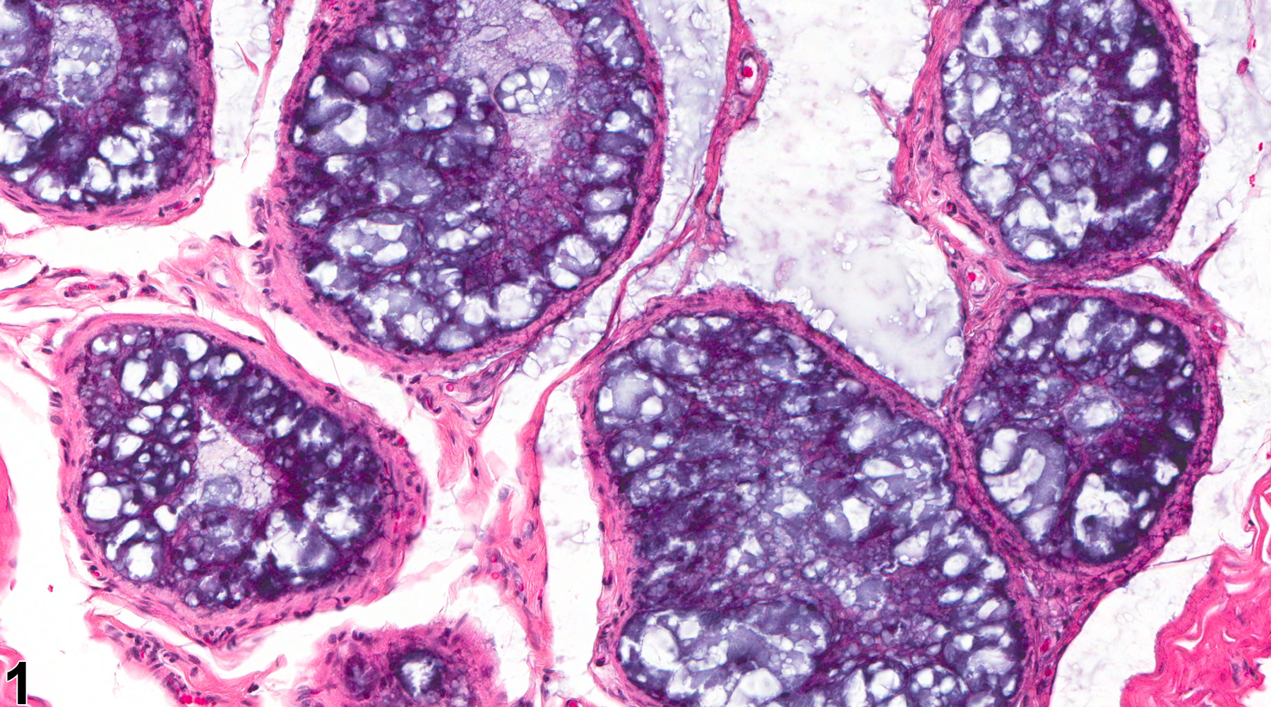
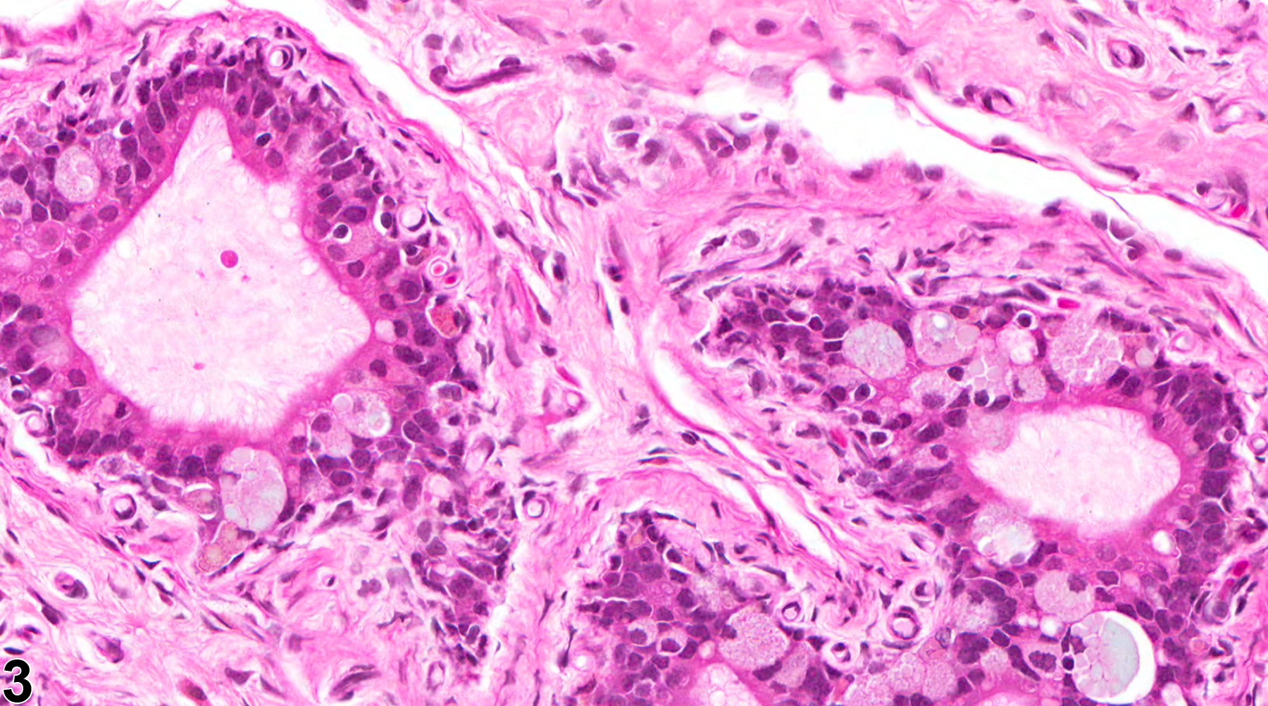
Epididymis, Epithelium - Vacuolation. Numerous macrovesicular cytoplasmic vacuoles are present in the ductal epithelium in a male F344/N rat from a chronic study.
Figure 1 of 3
Image of epithelial vacuolation in the epididymis from a  rat
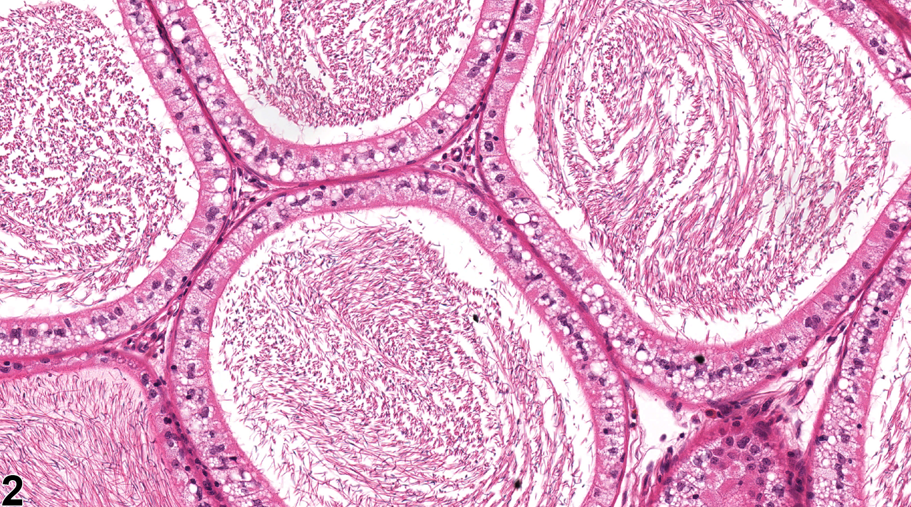
Epididymis, Epithelium - Vacuolation. Microvesicular vacuolation is present in the epididymal duct cells of a rat. (Photograph courtesy of D. Creasy.)
Figure 2 of 3
Image of epithelial vacuolation in the epididymis from a male F344/N rat in a chronic study
Epididymis, Epithelium - Clear cells. Clear cells in a male F344/N rat from a chronic study. Clear cells are a normal cell type responsible for endocytosis of luminal particulate matter and should not be confused with degenerative cytoplasmic vacuolation.
Figure 3 of 3
next arrow

comment:

The epididymal epithelium develops many different forms of vacuolation. They are often segmental and region specific. The vacuolation may be the result of a number of degenerative processes, including the accumulation of lipids, phospholipids (phospholipidosis), glycoproteins, or fluids. The vacuoles may be or large (macrovacuolation, Figure 1image opens in a pop-up window ) or small (microvacuolation, Figure 2image opens in a pop-up window ) and may appear empty or filled with basophilic or eosinophilic material. Basophilic vacuolation is a common age-related, background finding in a specific segment of the distal caput region of the rat ( Figure 1image opens in a pop-up window ). Large clear vacuoles are frequently seen as an incidental finding at the corpus cauda junction in the rat epididymis. Vacuolation of the epididymis can also be test article related and may be diffuse or segmental. Clear cells are a normal cell type of the epididymal epithelium involved in endocytosis of luminal particulate matter. They are often increased in number and prominence, particularly in the cauda, when there is increased cell debris in the epididymal lumen ( Figure 3image opens in a pop-up window ). Care should be taken to distinguish these normal clear cells from degenerative vacuolar changes. The distinction can be made since clear cells are filled with lysosomes that stain strongly positive with Periodic acid-Schiff.

recommendation:

Vacuolation of the epithelium should be recorded and graded and should be discussed in the pathology narrative if the incidence and/or severity appears to be related to chemical administration. The type and location of the vacuolation should be noted if it appears specific and noteworthy. Diagnosis should indicate if the change is bilateral. Correlation with disturbances in other male reproductive organs is recommended to aid interpretation.

references:

Boorman GA, Chapin RE, Mitsumori K. 1990. Testis and epididymis. In: Pathology of the Fischer Rat: Reference and Atlas (Boorman GA, Eustis SL, Elwell MR, Montgomery CA, MacKenzie WF, eds). Academic Press, San Diego, 405-418.

Creasy D, Bube A, de Rijk E, Kandori H, Kuwahara M, Masson R, Nolte T, Reams R, Regan K, Rehm S, Rogerson P, Whitney K. 2012. Proliferative and nonproliferative lesions of the rat and mouse male reproductive system. Toxicol Pathol 40:40S-121S.
Abstract: http://www.ncbi.nlm.nih.gov/pubmed/22949412

Haines DC, Chattopadhyay S, Ward JM. 2001. Pathology of aging B6;129 mice. Toxicol Pathol 29:653-661.
Abstract: http://www.ncbi.nlm.nih.gov/pubmed/11794381

Robaire B. 2002. Aging of the epididymis. In: The Epididymis: From Molecules to Clinical (Robaire B, Hinton BT, eds). New York, Kluwer/Plenum, 285-296.
Abstract: http://www.springer.com/medicine/urology/book/978-0-306-46684-7

Robaire B, Hinton BT, eds. 2002. The Epididymis: From Molecules to Clinical.
Abstract: http://www.springer.com/medicine/urology/book/978-0-306-46684-7

Rudmann DG, McNerney ME, Vandereide SL, Schemmer JK, Eversole RR, Vonderfecht SL. 2004. Epididymal and systemic phospholipidosis in rats and dogs treated with the dopamine D3 selective antagonist PNU-177864. Toxicol Pathol 32:326-332.
Abstract: http://www.ncbi.nlm.nih.gov/pubmed/15204974

Stebbins KE, Bond DM, Novilla MN, Reasor MJ. 2002. Spinosad insecticide: Subchronic and chronic toxicity and lack of carcinogenicity in CD-1 mice. Toxicol Sci 65:276-287.
Abstract: http://toxsci.oxfordjournals.org/content/65/2/276.abstract

Yano BL, Bond DM, Novilla MN, McFadden LG, Reasor MJ. 2002. Spinosad insecticide: Subchronic and chronic toxicity and lack of carcinogenicity in Fischer 344 rats. Toxicol Sci 65:288-298.
Abstract: http://toxsci.oxfordjournals.org/content/65/2/288.abstract