

Reproductive System, Male
Epididymis - Inflammation
Narrative
NTP studies have five standard categories of inflammation: acute, suppurative, chronic, chronic-active, and granulomatous. In acute inflammation, the predominant infiltrating cell is the neutrophil, though fewer macrophages and lymphocytes may also be present. There may also be evidence of edema or hyperemia. The neutrophil is also the predominant infiltrating cell type in suppurative inflammation, but they are aggregated, and many of them are degenerate (suppurative exudate). Cell debris from both the resident cell populations and infiltrating leukocytes, proteinaceous fluid containing fibrin, fewer macrophages, occasional lymphocytes or plasma cells, and, possibly, an infectious agent may also be present in the exudate. Grossly, these lesions would be characterized by the presence of pus. The tissue surrounding the exudate may have fibroblasts, fibrous connective tissue, and mixed inflammatory cells, depending on the chronicity of the lesion. Lymphocytes predominate in chronic inflammation. Lymphocytes also predominate in chronic-active inflammation, but there are also a significant number of neutrophils. Both lesions may contain macrophages. Granulomatous inflammation is another form of chronic inflammation, but this diagnosis requires the presence of a significant number of aggregated, large, activated macrophages, epithelioid macrophages, or multinucleated giant cells.
Atanassova N, McKinnell C, Fisher J, Sharpe RM. 2005. Neonatal treatment of rats with diethylstilbestrol (DES) induces stromal-epithelial abnormalities of the vas deferens and cauda epididymis in adulthood following delayed basal cell development. Reproduction 129:589-601.
Abstract: http://www.ncbi.nlm.nih.gov/pubmed/15855622Chapin RE, White RD, Morgan KT, Bus JS. 1984. Studies of lesions induced in the testis and epididymis of F-344 rats by inhaled methyl chloride. Toxicol Appl Pharmacol 76:328-343.
Abstract: http://www.ncbi.nlm.nih.gov/pubmed/6388032Chellman GJ, Morgan KT, Bus JS, Working PK. 1986. Inhibition of methyl chloride toxicity in male F-344 rats by the anti-inflammatory agent BW-755C. Toxicol Appl Pharmacol 85:365-379.
Abstract: http://www.ncbi.nlm.nih.gov/pubmed/3094195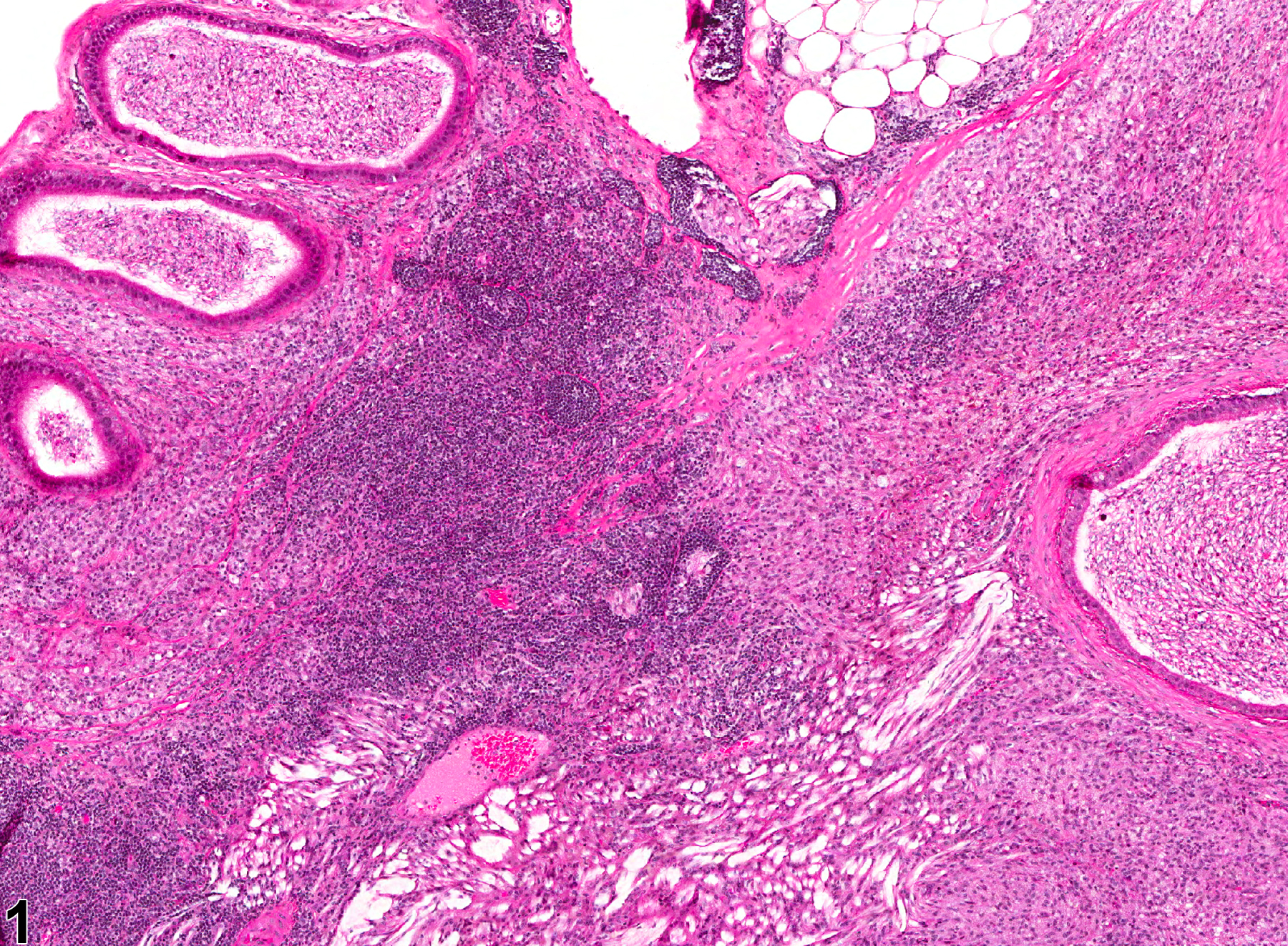
Epididymis - Inflammation. Chronic inflammation with lymphoid aggregates in the epididymis in a male B6C3F1 mouse from a chronic study.
All Images

Epididymis - Inflammation. Chronic inflammation with lymphoid aggregates in the epididymis in a male B6C3F1 mouse from a chronic study.

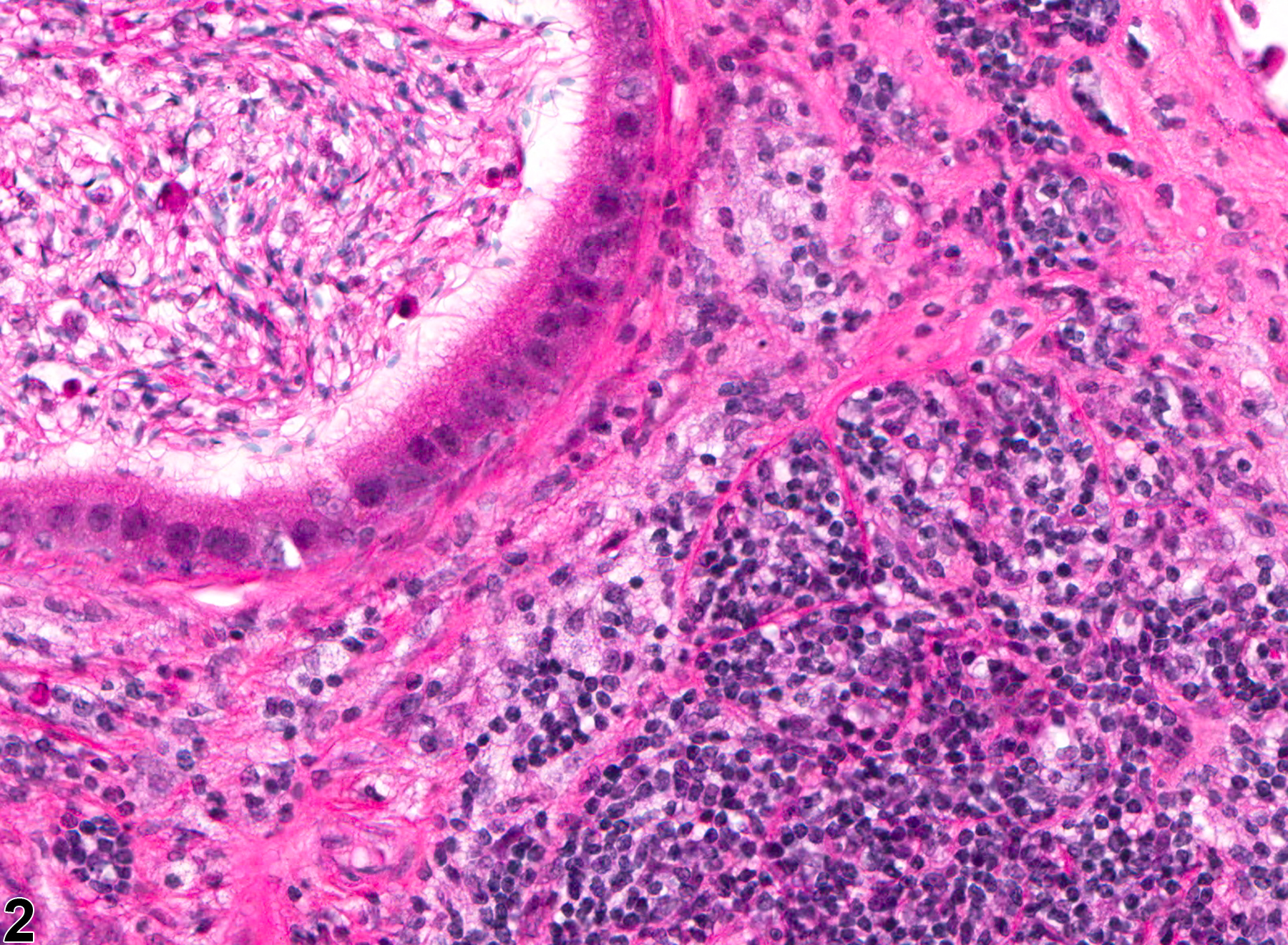
Epididymis - Inflammation. Higher magnification of Figure 1. Chronic inflammation with lymphoid aggregates in the epididymis in a male B6C3F1 mouse from a chronic study.
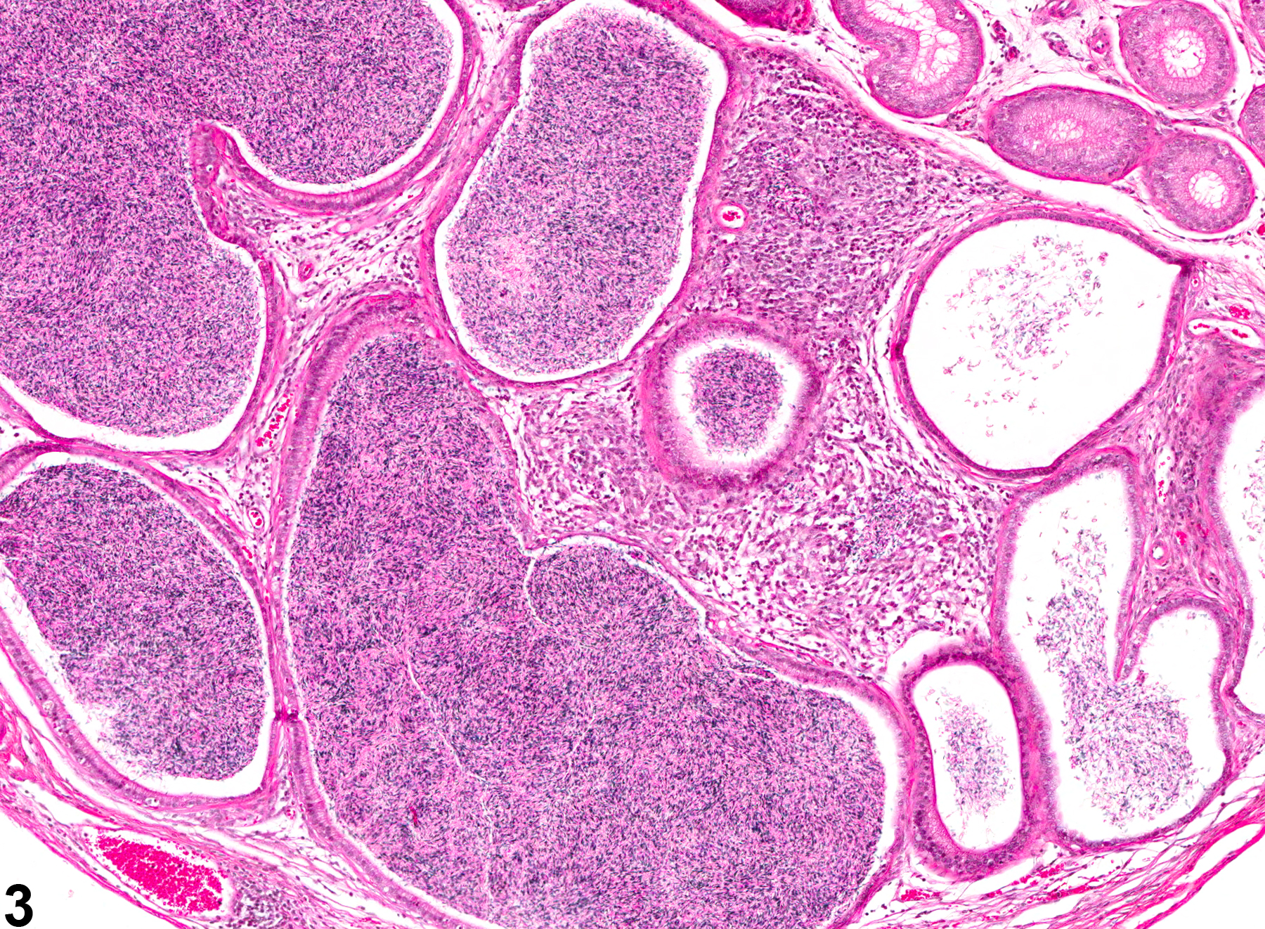
Epididymis - Inflammation. A primarily mononuclear infiltration has enlarged the interstitium between duct profiles in a male B6C3F1 mouse from a chronic study.
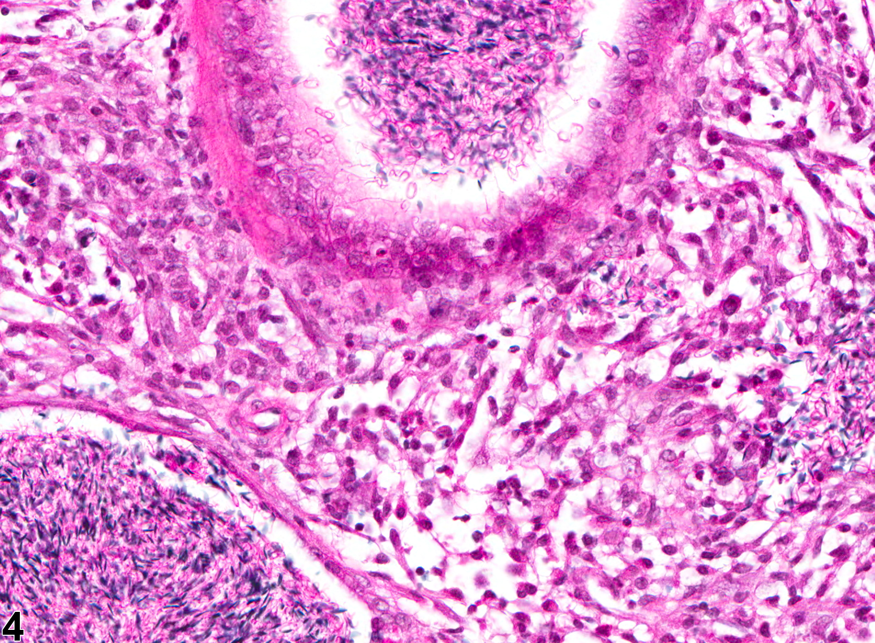
Epididymis - Inflammation. Higher magnification of Figure 3 showing mononuclear inflammatory cells between duct profiles in a male B6C3F1 mouse from a chronic study.