U.S. flag

An official website of the United States government

Dot gov

The .gov means it's official.
Federal government websites often end in .gov or .mil. Before sharing sensitive information, make sure you're on a federal government site.

Https

The site is secure.
The https:// ensures that you are connecting to the official website and that any information you provide is encrypted and transmitted securely.

Skip to Main Navigation
Skip to Page Content
Skip to Atlas Navigation

Epididymis - Spermatocele

Image of spermatocele in the epididymis from a male B6C3F1 mouse in a subchronic study
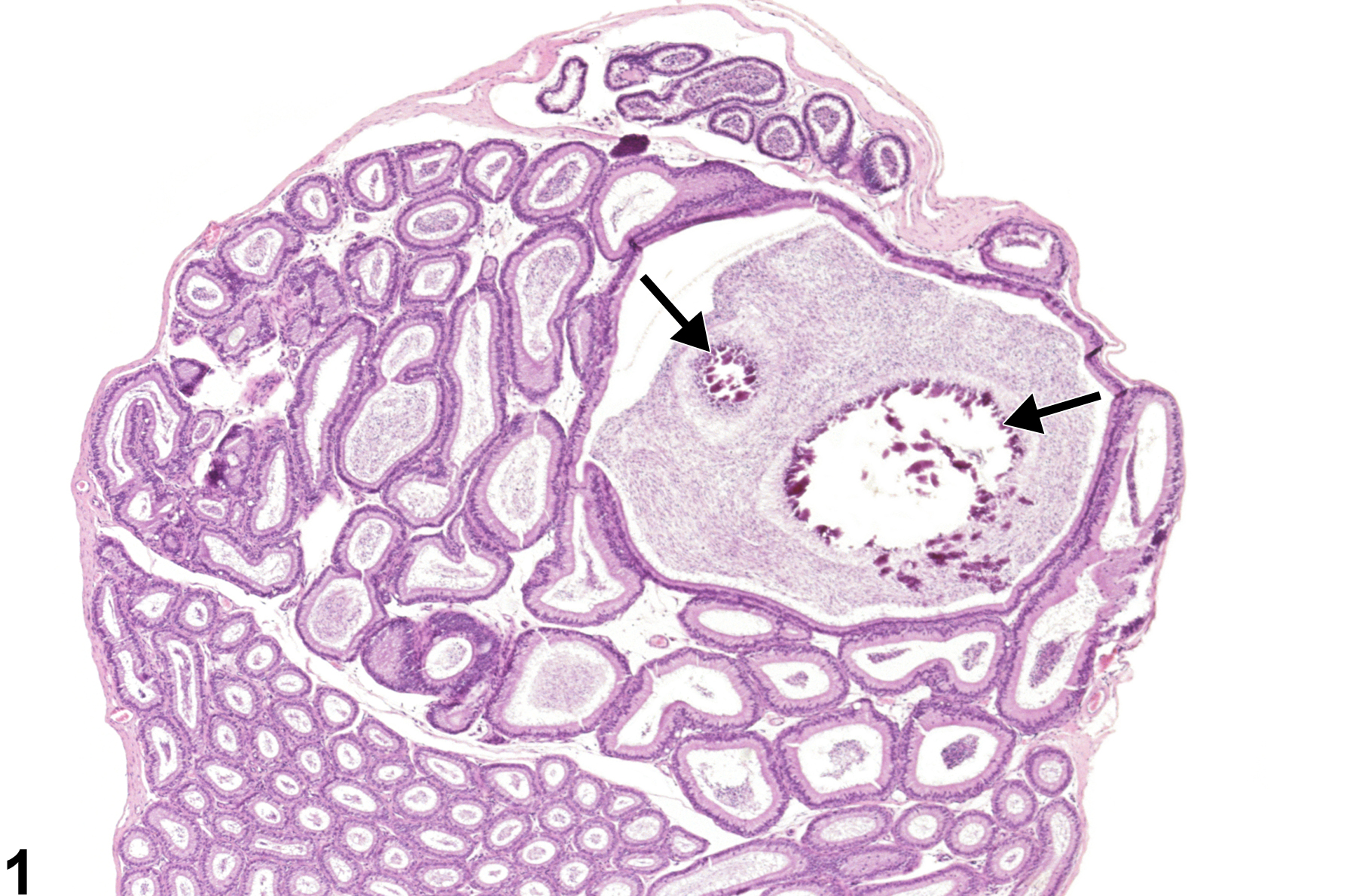
Epididymis - Spermatocele. A sperm-filled dilated duct in the head of the epididymis, with focal areas of mineralization (arrows), in a male B6C3F1 mouse from a subchronic study.
Figure 1 of 1
next arrow

comment:

Spermatoceles are cystic dilations of the epididymal duct (three or more times the diameter of the normal duct), which may be lined by hypertrophic or normal epithelium. They are most commonly seen in the initial segment or the caput region of the mouse epididymis (Figure 1) and are filled with varying amounts of sperm. Focal areas of mineralization are present within this spermatocele (arrows, Figure 1image opens in a pop-up window ). Spermatoceles generally occur as background, incidental lesions but have been reported in association with exposure to some chemicals. They are distinguished from sperm granuloma by the absence of an inflammatory response.

recommendation:

Spermatoceles should be recorded as present and discussed in the pathology narrative if the incidence appears to be related to chemical administration. If present in both epididymides, the diagnosis should be qualified as bilateral. It is not necessary to grade spermatoceles.

references:

Cooper ERA, Jackson H. 1973. Chemically induced sperm retention cysts in the rat. J Reprod Fertil 34:445-449.
Abstract: http://www.popline.org/node/491769

Creasy D, Bube A, de Rijk E, Kandori H, Kuwahara M, Masson R, Nolte T, Reams R, Regan K, Rehm S, Rogerson P, Whitney K. 2012. Proliferative and nonproliferative lesions of the rat and mouse male reproductive system. Toxicol Pathol 40:40S-121S.
Abstract: http://www.ncbi.nlm.nih.gov/pubmed/22949412

Frith CH, Ward JM. 1988. Color Atlas of Neoplastic and Non-neoplastic Lesions in Aging Mice. Elsevier, Amsterdam.
Abstract: http://www.informatics.jax.org/frithbook/

Hess RA. 1998. Effects of environmental toxicants on the efferent ducts, epididymis and fertility. J Reprod Fertil Suppl 53:247-259.
Abstract: http://www.ncbi.nlm.nih.gov/pubmed/10645284

Radovsky A, Mitsumori K, Chapin RE. 1999. Male reproductive tract. In: Pathology of the Mouse: Reference and Atlas (Maronpot RR, Boorman GA, Gaul BW, eds). Cache River Press, Vienna, IL, 381-407.
Abstract: http://www.cacheriverpress.com/books/pathmouse.htm