U.S. flag

An official website of the United States government

Dot gov

The .gov means it's official.
Federal government websites often end in .gov or .mil. Before sharing sensitive information, make sure you're on a federal government site.

Https

The site is secure.
The https:// ensures that you are connecting to the official website and that any information you provide is encrypted and transmitted securely.

Skip to Main Navigation
Skip to Page Content
Skip to Atlas Navigation

Prostate, Acinus - Cyst(s), Mucinous

Image of acinar mucinous cyst in the prostate from a male F344/N rat in a chronic study
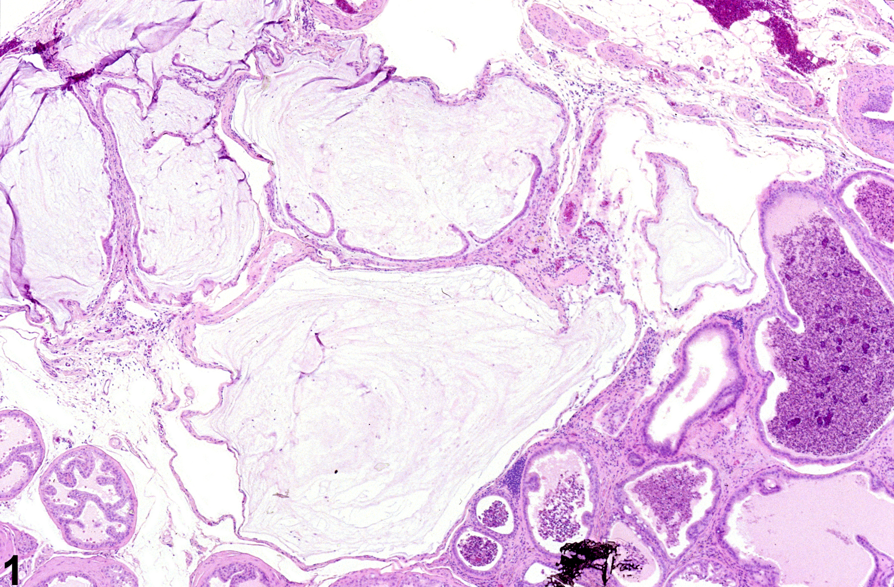
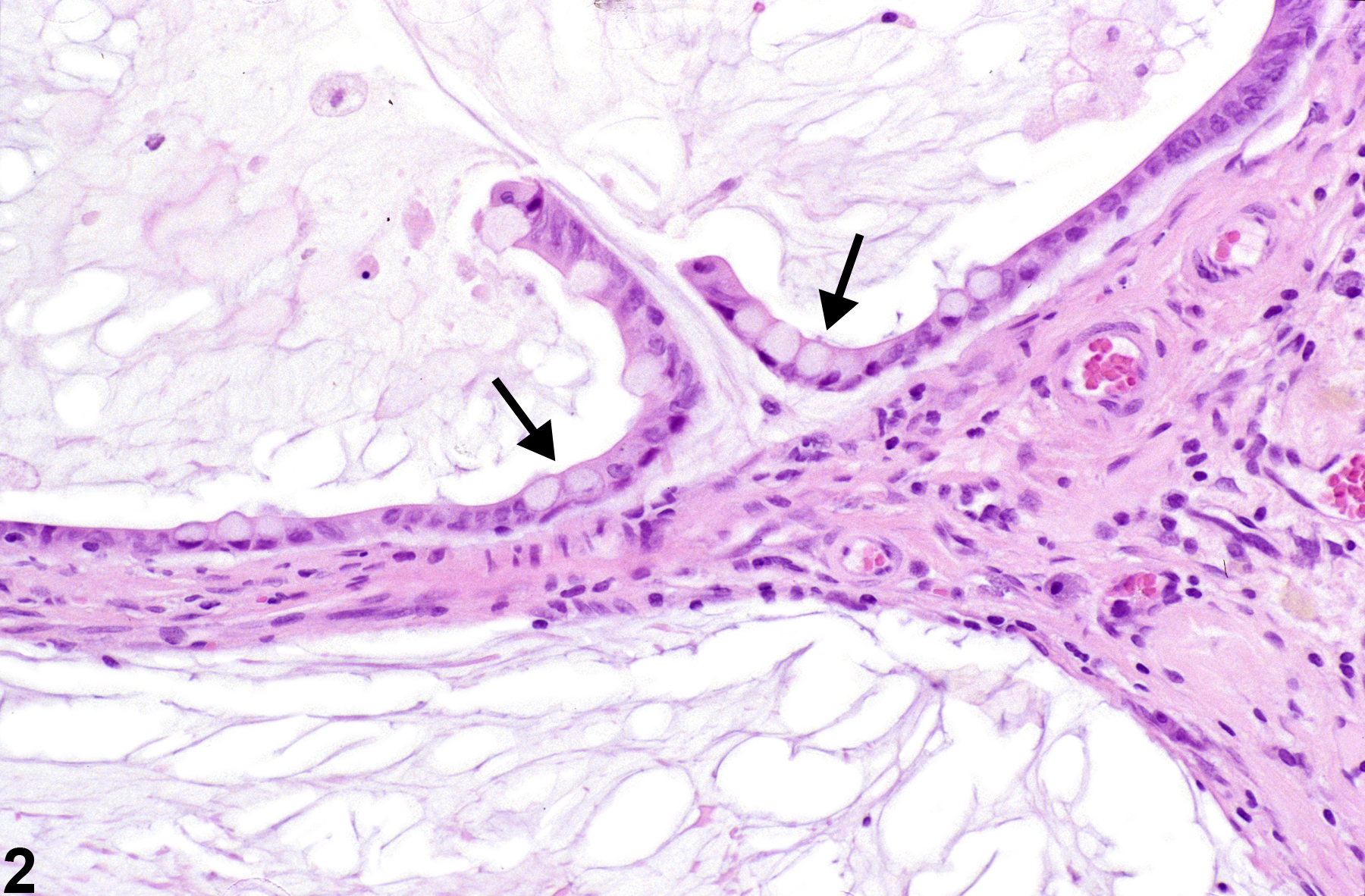
Prostate, Acinus - Cyst, Mucinous. Mucinous cyst in a male F344/N rat from a chronic study.
Figure 1 of 2
Image of acinar mucinous cyst in the prostate from a male F344/N rat in a chronic study
Prostate, Acinus - Cyst, Mucinous. Higher magnification of Figure 1. Mucinous cyst in a male F344/N rat from a chronic study. Goblet cells are present in the lining epithelium of the mucinous cysts (arrows).
Figure 2 of 2
next arrow

comment:

Although mucinous cysts are a type of prostate duct dilation, the condition has recently been seen in studies and warrants a separate diagnosis. Mucinous cysts ( Figure 1image opens in a pop-up window and Figure 2image opens in a pop-up window ) have been documented in a small number of treated F344 rats in emodin and anthraquinone chronic studies. Goblet cells are present in the lining epithelium of the mucinous cysts ( Figure 2image opens in a pop-up window , arrows).

recommendation:

Prostatic mucinous cysts represent a distinct entity that presently requires a separate diagnosis to distinguish it from acinar dilation. Their occurrence should be graded, and bilateral involvement should be documented if present, with severity based on the more severely affected lobe. When possible, the affected prostate lobe should be identified.

references:

Boorman GA, Elwell MR, Mitsumori K. 1990. Male accessory sex glands, penis, and scrotum. In: Pathology of the Fischer Rat: Reference and Atlas (Boorman GA, Eustis SL, Elwell MR, Montgomery CA, MacKenzie WF, eds). Academic Press, San Diego, 419-428.
Abstract: http://www.ncbi.nlm.nih.gov/nlmcatalog/9002563

Bosland MC. 1992. Lesions in the male accessory glands and penis. In: Pathobiology of the Aging Rat, Vol 1 (Mohr U, Dungworth DL, Capen CC, eds). ILSI Press, Washington, DC, 443-467.
Abstract: http://catalog.hathitrust.org/Record/008994685

Creasy D, Bube A, de Rijk E, Kandori H, Kuwahara M, Masson R, Nolte T, Reams R, Regan K, Rehm S, Rogerson P, Whitney K. 2012. Proliferative and nonproliferative lesions of the rat and mouse male reproductive system. Toxicol Pathol 40:40S-121S.
Abstract: http://www.ncbi.nlm.nih.gov/pubmed/22949412

Gordon LR, Majka JA, Boorman GA. 1996. Spontaneous nonneoplastic and neoplastic lesions and experimentally induced neoplasms of the testes and accessory sex glands. In: Pathobiology of the Aging Mouse, Vol 1 (Mohr U, Dungworth DL, Capen CC, Carlton WW, Sundberg JP, Ward JM, eds). ILSI Press, Washington, DC, 421-441.
Abstract: http://catalog.hathitrust.org/Record/008994685

Greaves P. 2007. Male genital tract. In: Histopathology of Preclinical Toxicity Studies: Interpretation and Relevance in Drug Safety Evaluation. 3rd ed. Academic Press, San Diego, 661-716.
Abstract: http://www.sciencedirect.com/science/book/9780444527714