

Reproductive System, Male
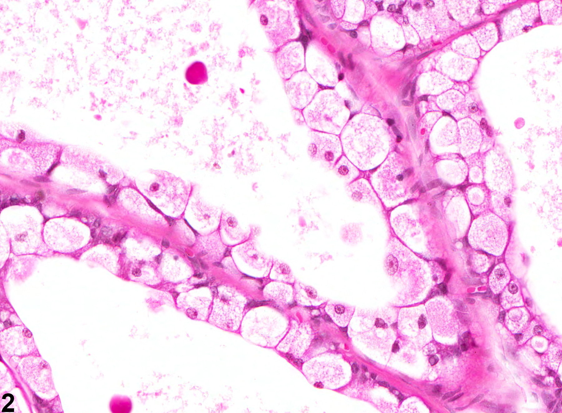
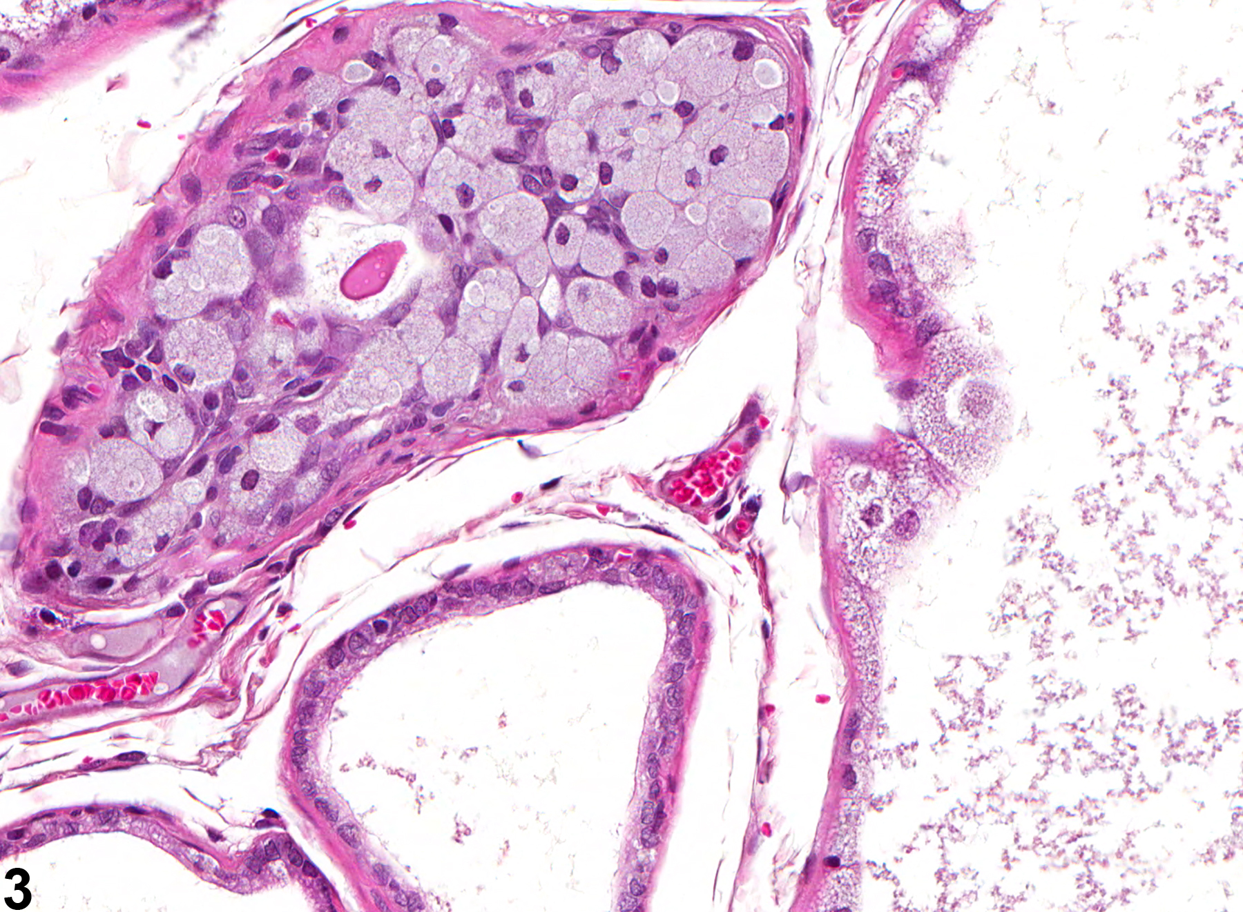
Prostate, Epithelium - Degeneration
Narrative
Bosland MC. 1992. Lesions in the male accessory glands and penis. In: Pathobiology of the Aging Rat, Vol 1 (Mohr U, Dungworth DL, Capen CC, eds). ILSI Press, Washington, DC, 443-467.
Abstract: http://catalog.hathitrust.org/Record/008994685Gordon LR, Majka JA, Boorman GA. 1996. Spontaneous nonneoplastic and neoplastic lesions and experimentally induced neoplasms of the testes and accessory sex glands. In: Pathobiology of the Aging Mouse, Vol 1 (Mohr U, Dungworth DL, Capen CC, Carlton WW, Sundberg JP, Ward JM, eds). ILSI Press, Washington, DC, 421-441.
Abstract: http://catalog.hathitrust.org/Record/008994685Greaves P. 2007. Male genital tract. In: Histopathology of Preclinical Toxicity Studies: Interpretation and Relevance in Drug Safety Evaluation. 3rd ed. Academic Press, San Diego, 661-716.
Abstract: http://www.sciencedirect.com/science/book/9780444527714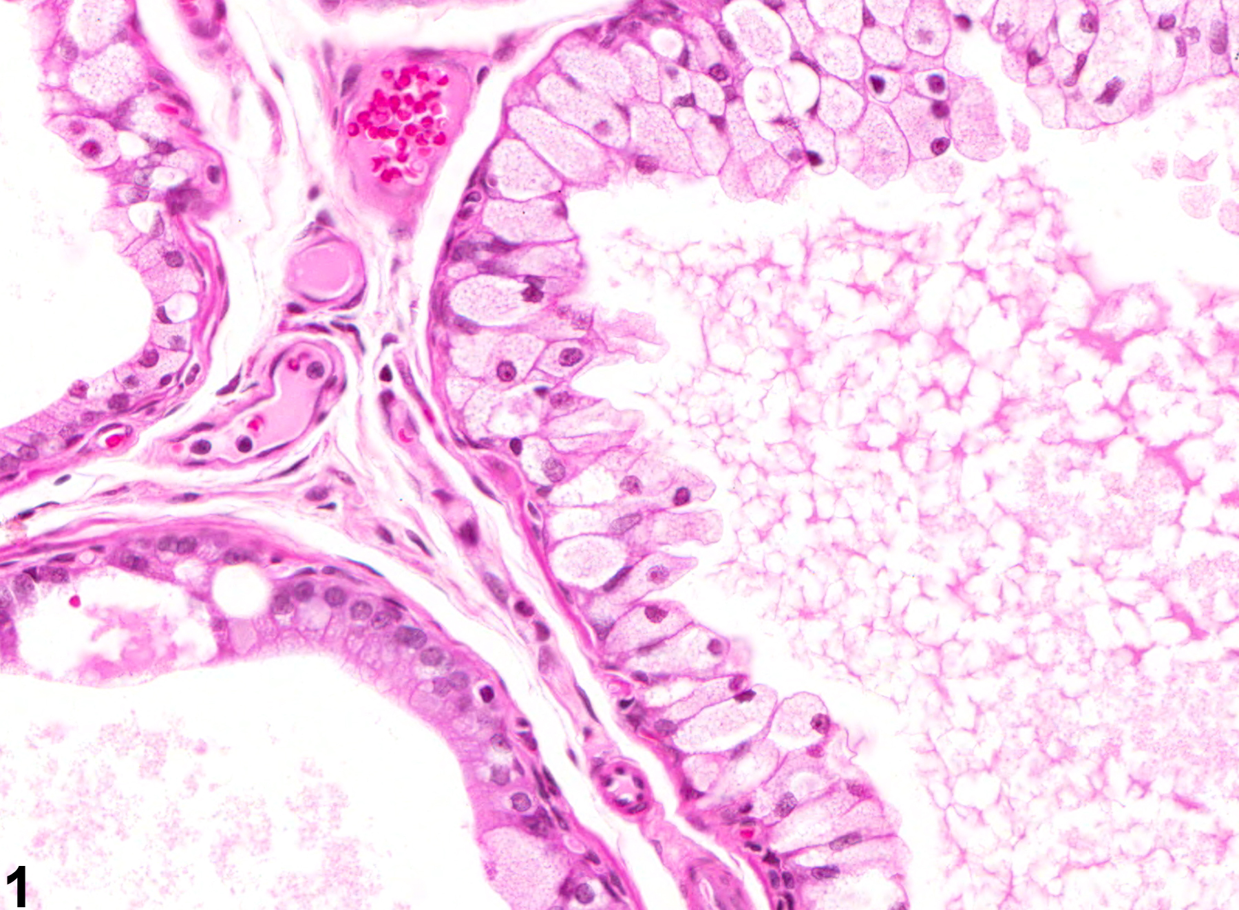
Prostate, Epithelium - Degeneration. Degeneration of the epithelium in the prostate in a male F344/N rat from a chronic study.
All Images

Prostate, Epithelium - Degeneration. Degeneration of the epithelium in the prostate in a male F344/N rat from a chronic study.

Prostate, Epithelium - Degeneration. Degeneration of the epithelium in the prostate in a male F344/N rat from a chronic study.

Prostate, Epithelium - Degeneration. Degeneration of the epithelium in the prostate in a male F344/N rat from a chronic study.