

Reproductive System, Male
Seminal Vesicle - Atrophy
Narrative
Boorman GA, Elwell MR, Mitsumori K. 1990. Male accessory sex glands, penis, and scrotum. In: Pathology of the Fischer Rat: Reference and Atlas (Boorman GA, Eustis SL, Elwell MR, Montgomery CA, MacKenzie WF, eds). Academic Press, San Diego, 419-428.
Abstract: http://www.ncbi.nlm.nih.gov/nlmcatalog/9002563Bosland MC. 1992. Lesions in the male accessory glands and penis. In: Pathobiology of the Aging Rat, Vol 1 (Mohr U, Dungworth DL, Capen CC, eds). ILSI Press, Washington, DC, 443-467.
Abstract: http://catalog.hathitrust.org/Record/008994685Comereski CR, Pedan WM, Davidson TJ, Warner GL, Hirth RS, Frantz JD. 1994. BR96-doxorubicin conjugate (BMS-182248) versus doxorubicin: A comparative toxicity assessment in rats. Toxicol Pathol 22:473-488.
Abstract: http://www.ncbi.nlm.nih.gov/pubmed/7899776Gayton F, Bellido C, Aguilar R, Lucena MC. 1986. Morphometric analysis of the rat ventral prostate and seminal vesicles during prepubertal development: Effects of neonatal treatment with estrogen. Biol Reprod 35:219-225.
Abstract: http://www.ncbi.nlm.nih.gov/pubmed/3741952Gordon LR, Majka JA, Boorman GA. 1996. Spontaneous nonneoplastic and neoplastic lesions and experimentally induced neoplasms of the testes and accessory sex glands. In: Pathobiology of the Aging Mouse, Vol 1 (Mohr U, Dungworth DL, Capen CC, Carlton WW, Sundberg JP, Ward JM, eds). ILSI Press, Washington, DC, 421-441.
Abstract: http://catalog.hathitrust.org/Record/008994685Greaves P. 2007. Male genital tract. In: Histopathology of Preclinical Toxicity Studies: Interpretation and Relevance in Drug Safety Evaluation. 3rd ed. Academic Press, San Diego, 661-716.
Abstract: http://www.sciencedirect.com/science/book/9780444527714Suwa T, Nyska A, Peckham JC, Hailey JR, Mahler JF, Haseman JK, Maronpot RR. 2001. A retrospective analysis of background lesions and tissue accountability for male accessory sex organs in Fischer-344 rats. Toxicol Pathol 29(4):467-478.
Abstract: http://www.ncbi.nlm.nih.gov/pubmed/11560252Suwa T, Nyska A, Haseman JK, Mahler JF, Maronpot RR. 2002. Spontaneous lesions in control B6C3F1 mice and recommended sectioning of male accessory sex organs. Toxicol Pathol 30(2):228-234.
Abstract: http://www.ncbi.nlm.nih.gov/pubmed/11950166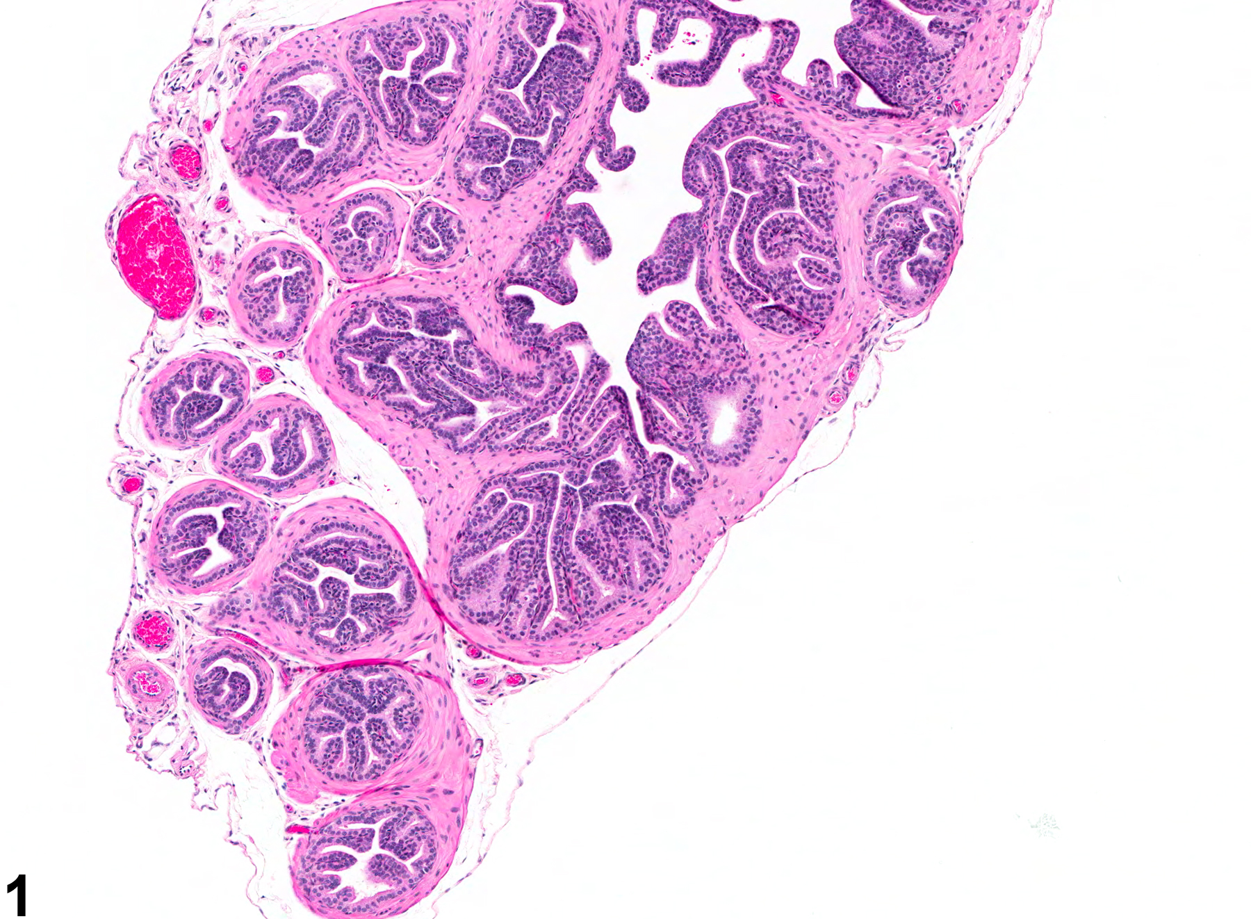
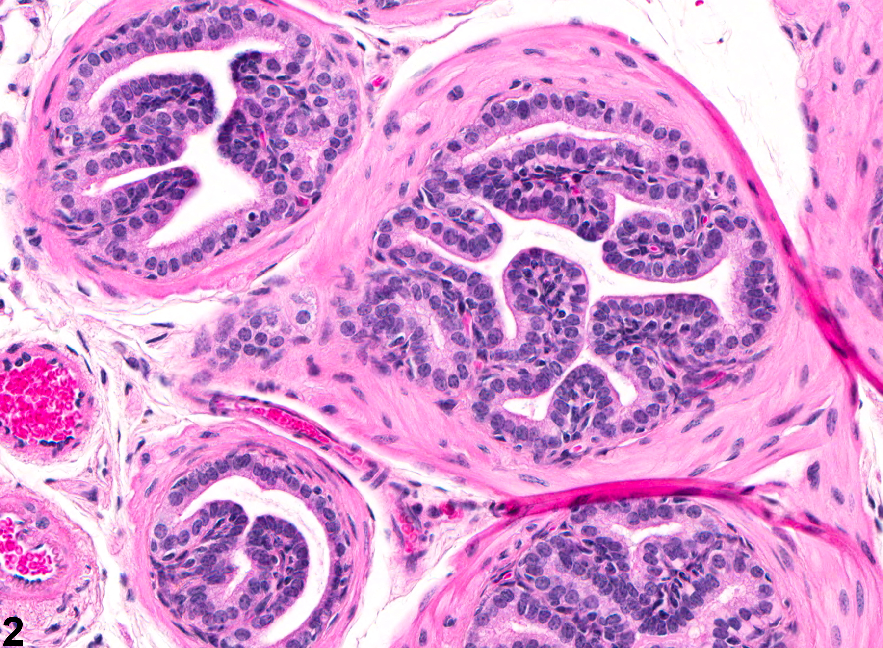
Seminal Vesicle - Atrophy. Atrophy of the seminal vesicle in a male B6C3F1 mouse from a subchronic study.
All Images

Seminal Vesicle - Atrophy. Atrophy of the seminal vesicle in a male B6C3F1 mouse from a subchronic study.

Seminal Vesicle - Atrophy. Higher magnification of Figure 1. Atrophy of the seminal vesicle in a male B6C3F1 mouse from a subchronic study.

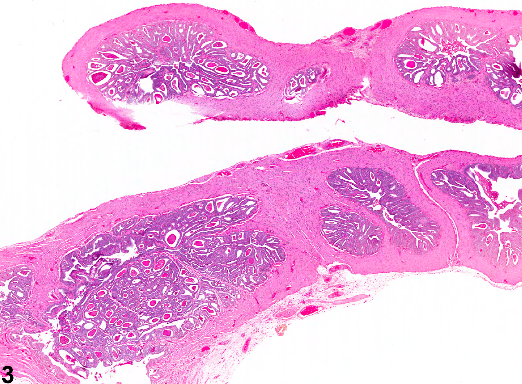
Seminal Vesicle - Atrophy. Atrophy of the seminal vesicle in a male F344/N rat from a subchronic study.

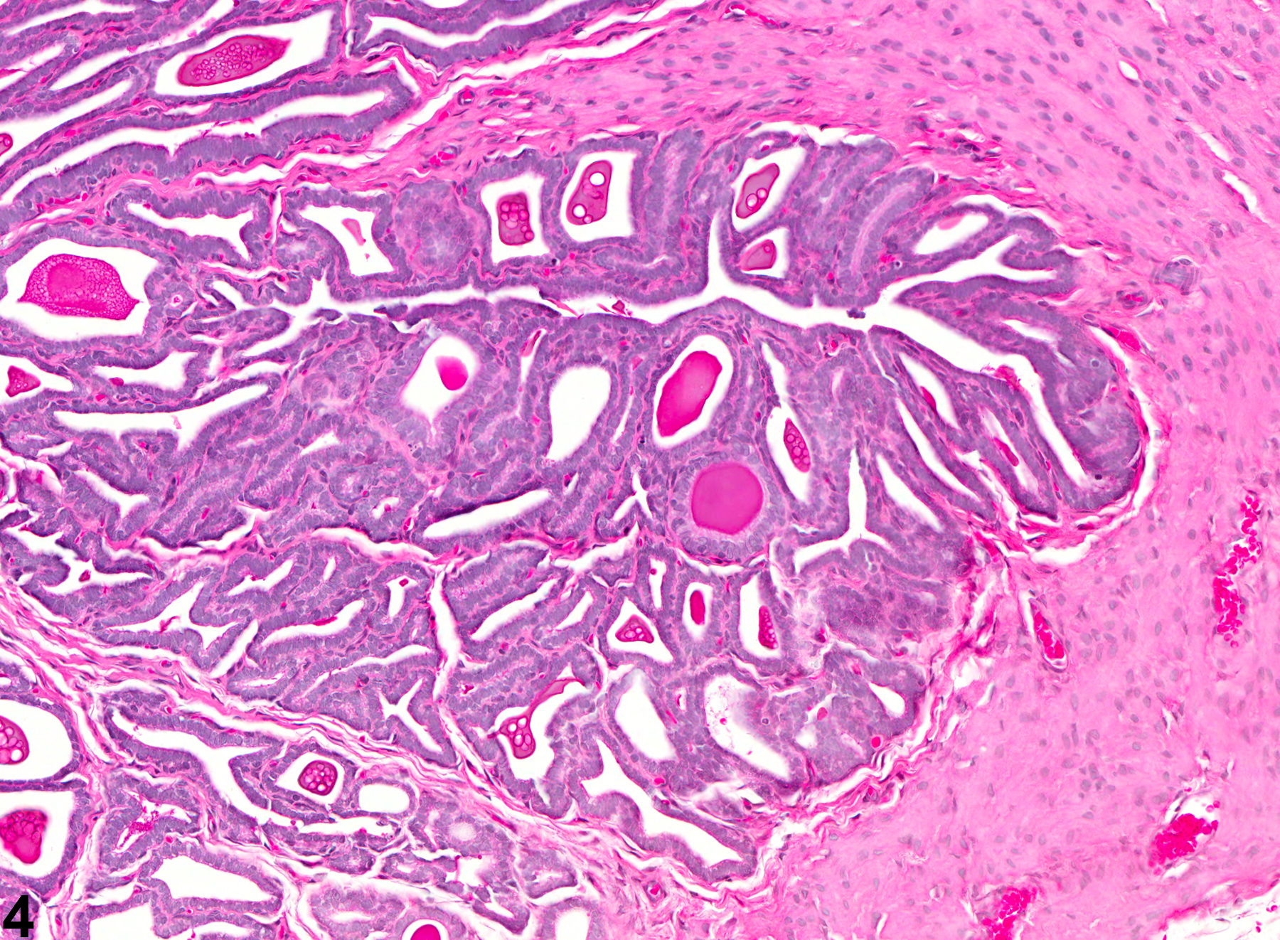
Seminal Vesicle - Atrophy. Higher magnification of Figure 3. Atrophy of the seminal vesicle in a male F344/N rat from a subchronic study.