

Reproductive System, Male
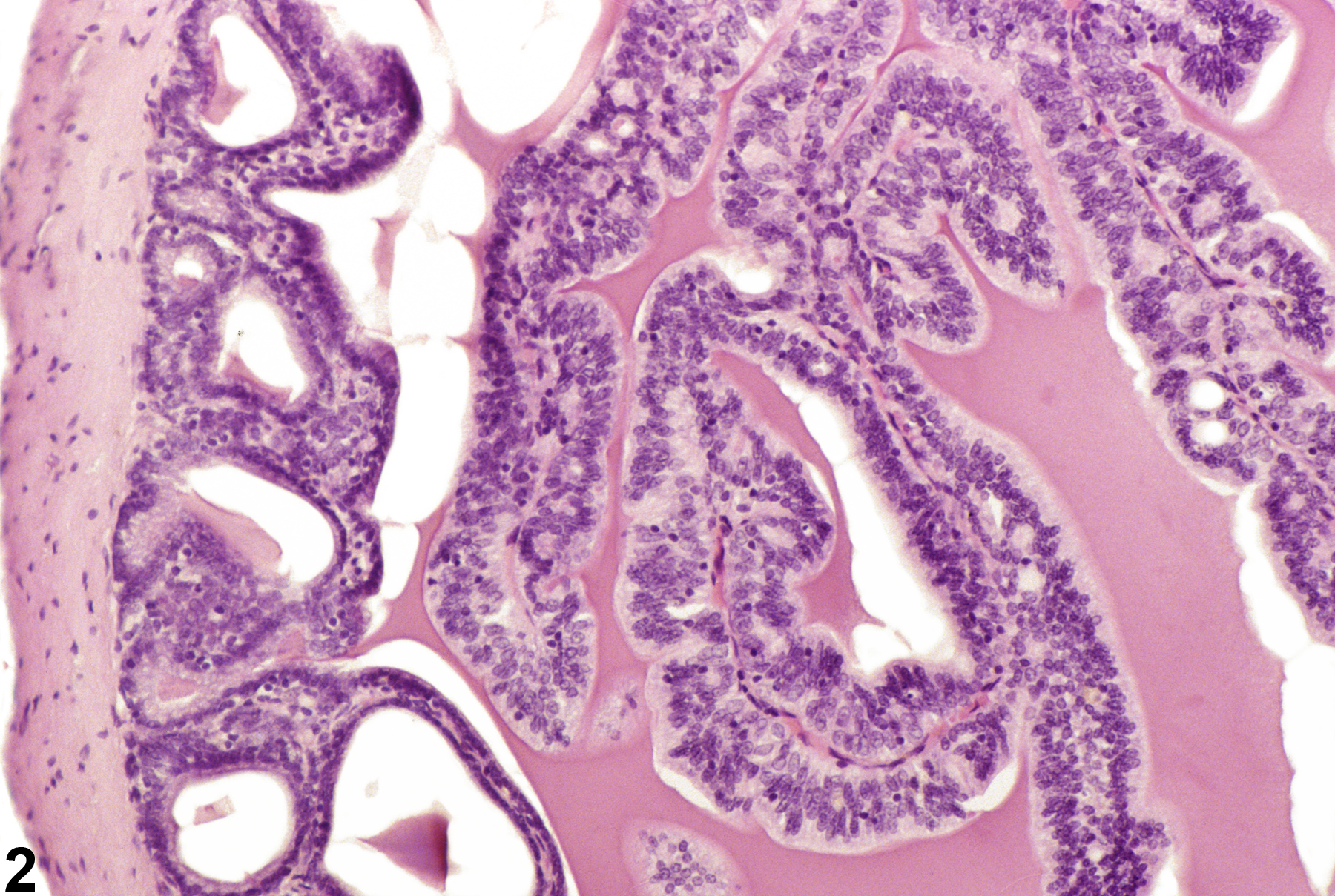
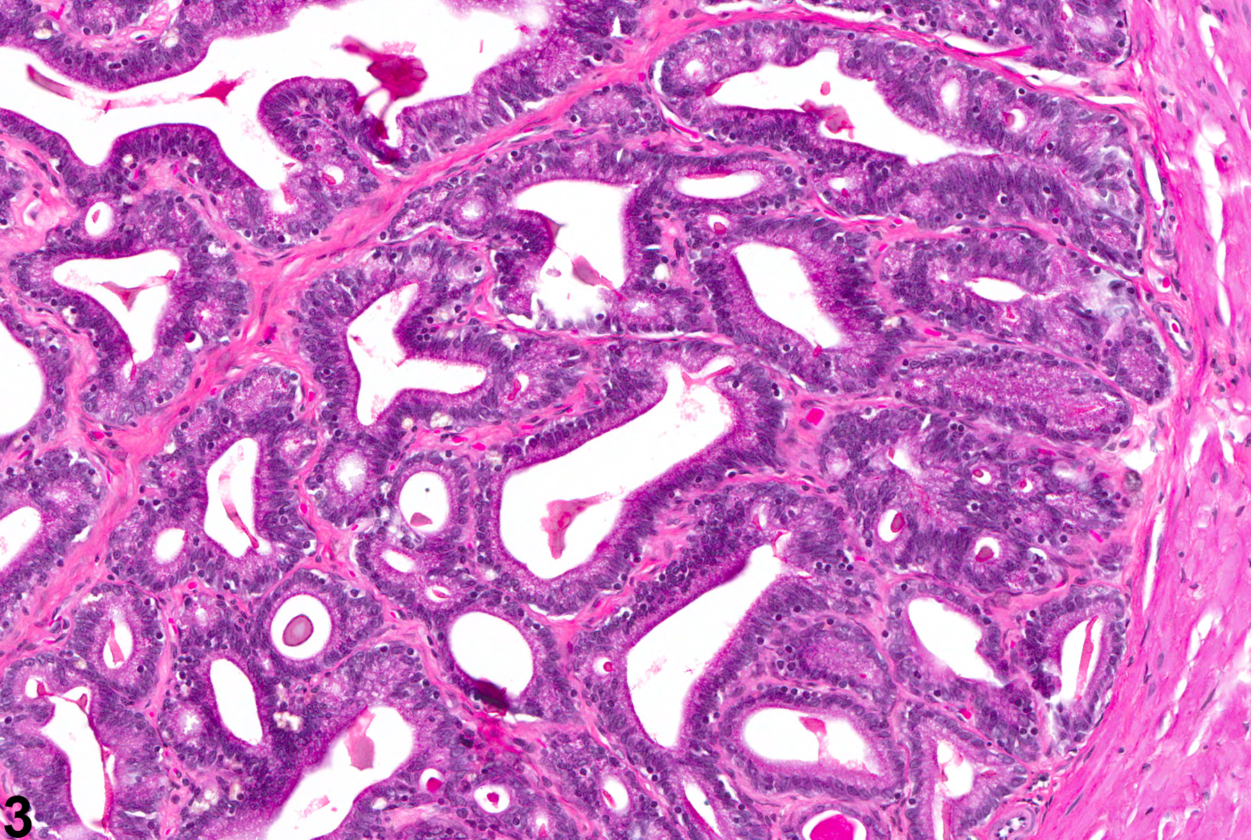
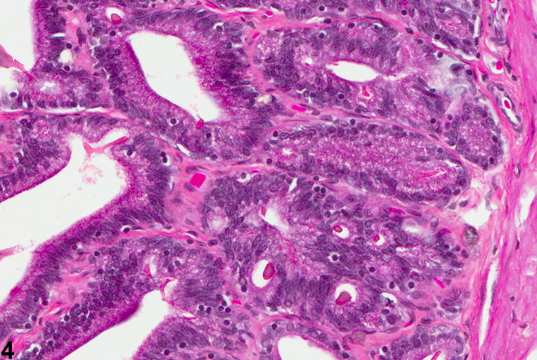
Seminal Vesicle, Epithelium - Hyperplasia
Narrative
Boorman GA, Elwell MR, Mitsumori K. 1990. Male accessory sex glands, penis, and scrotum. In: Pathology of the Fischer Rat: Reference and Atlas (Boorman GA, Eustis SL, Elwell MR, Montgomery CA, MacKenzie WF, eds). Academic Press, San Diego, 419-428.
Abstract: http://www.ncbi.nlm.nih.gov/nlmcatalog/9002563Bosland MC. 1992. Lesions in the male accessory glands and penis. In: Pathobiology of the Aging Rat, Vol 1 (Mohr U, Dungworth DL, Capen CC, eds). ILSI Press, Washington, DC, 443-467.
Abstract: http://catalog.hathitrust.org/Record/008994685Gordon LR, Majka JA, Boorman GA. 1996. Spontaneous nonneoplastic and neoplastic lesions and experimentally induced neoplasms of the testes and accessory sex glands. In: Pathobiology of the Aging Mouse, Vol 1 (Mohr U, Dungworth DL, Capen CC, Carlton WW, Sundberg JP, Ward JM, eds). ILSI Press, Washington, DC, 421-441.
Abstract: http://catalog.hathitrust.org/Record/008994685Radovsky A, Mitsumori K, Chapin RE. 1999. Male reproductive tract. In: Pathology of the Mouse: Reference and Atlas (Maronpot RR, Boorman GA, Gaul BW, eds). Cache River Press, Vienna, IL, 381-407.
Suwa T, Nyska A, Peckham JC, Hailey JR, Mahler JF, Haseman JK, Maronpot RR. 2001. A retrospective analysis of background lesions and tissue accountability for male accessory sex organs in Fischer-344 rats. Toxicol Pathol 29(4):467-478.
Abstract: http://www.ncbi.nlm.nih.gov/pubmed/11560252Suwa T, Nyska A, Haseman JK, Mahler JF, Maronpot RR. 2002. Spontaneous lesions in control B6C3F1 mice and recommended sectioning of male accessory sex organs. Toxicol Pathol 30(2):228-234.
Abstract: http://www.ncbi.nlm.nih.gov/pubmed/11950166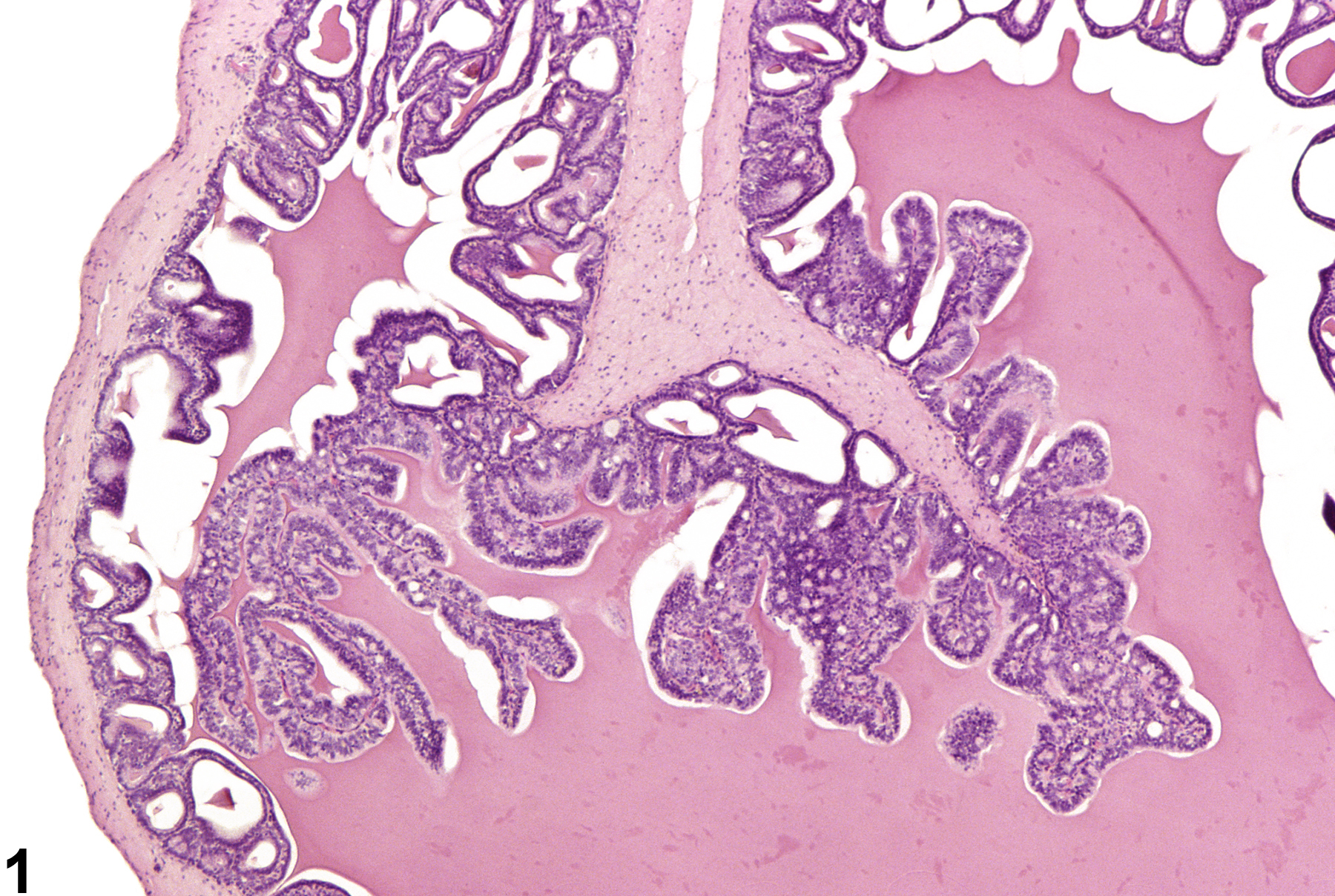
Seminal Vesicle, Epithelium - Hyperplasia. Hyperplasia of seminal vesicle epithelium in a male F344/N rat from a chronic study.
All Images

Seminal Vesicle, Epithelium - Hyperplasia. Hyperplasia of seminal vesicle epithelium in a male F344/N rat from a chronic study.

Seminal Vesicle, Epithelium - Hyperplasia. Higher magnification of Figure 1. Hyperplasia of seminal vesicle epithelium in a male F344/N rat from a chronic study.

Seminal Vesicle, Epithelium - Hyperplasia. Hyperplasia of seminal vesicle epithelium in a male F344/N rat from a chronic study.

Seminal Vesicle, Epithelium - Hyperplasia. Higher magnification of Figure 3. Hyperplasia of seminal vesicle epithelium in a male F344/N rat from a chronic study.