

Reproductive System, Male
Testis - Amyloid
Narrative
Gordon LR, Majka JA, Boorman GA. 1996. Spontaneous nonneoplastic and neoplastic lesions and experimentally induced neoplasms of the testes and accessory sex glands. In: Pathobiology of the Aging Mouse, Vol 1 (Mohr U, Dungworth DL, Capen CC, Carlton WW, Sundberg JP, Ward JM, eds). ILSI Press, Washington, DC, 421-441.
Abstract: http://catalog.hathitrust.org/Record/008994685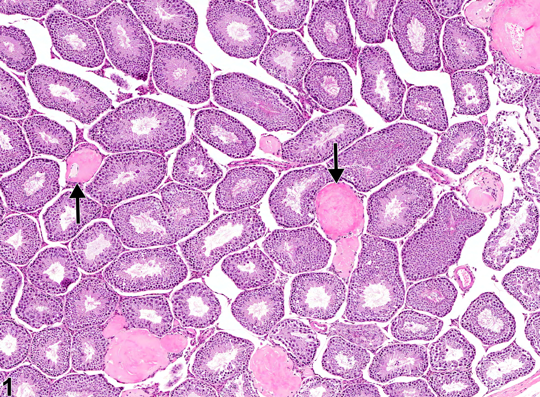
Testis - Amyloid in a male B6C3F1 mouse from a chronic study. There are perivascular accumulations of hemogenous, eosinophilic material in the interstitium (arrows).
All Images

Testis - Amyloid in a male B6C3F1 mouse from a chronic study. There are perivascular accumulations of hemogenous, eosinophilic material in the interstitium (arrows).

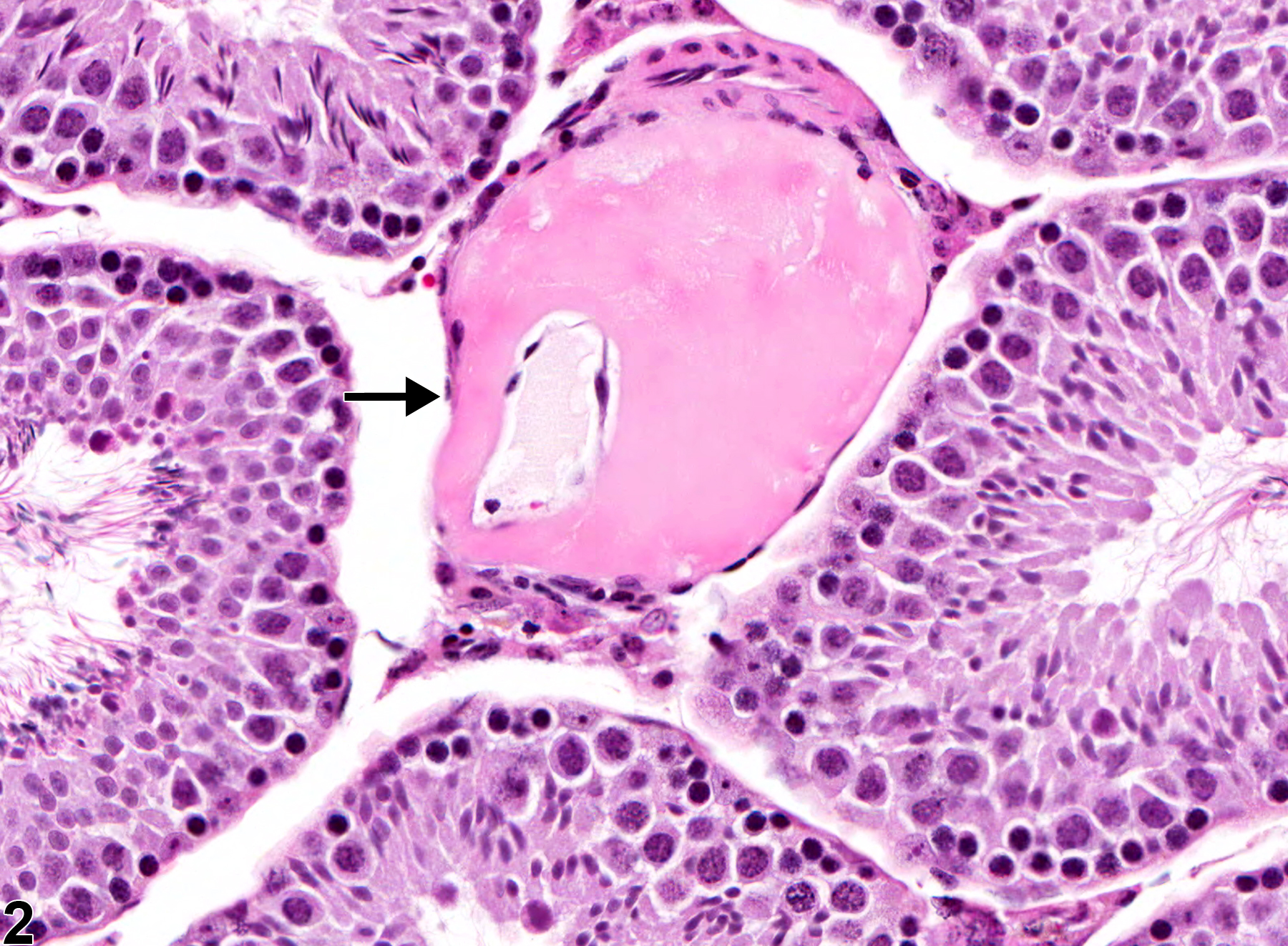
Testis - Amyloid in a male B6C3F1 mouse from a chronic study. Higher magnification of Figure 1 with perivascular accumulation of amyloid (arrow).

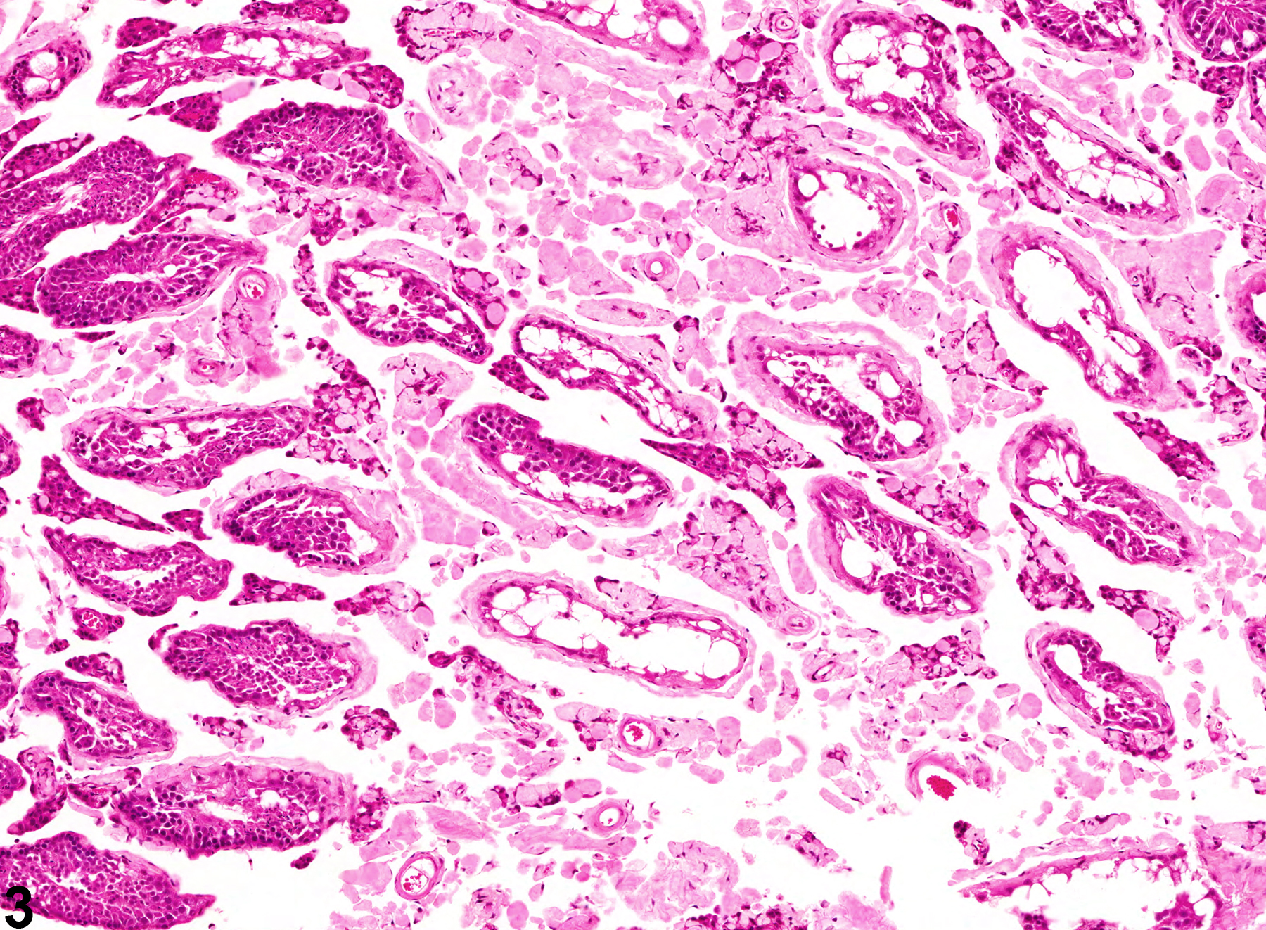
Testis - Amyloid in a male Swiss CD-1 mouse from a chronic study. Widespread accumulation of amyloid with secondary tubular degeneration and necrosis.

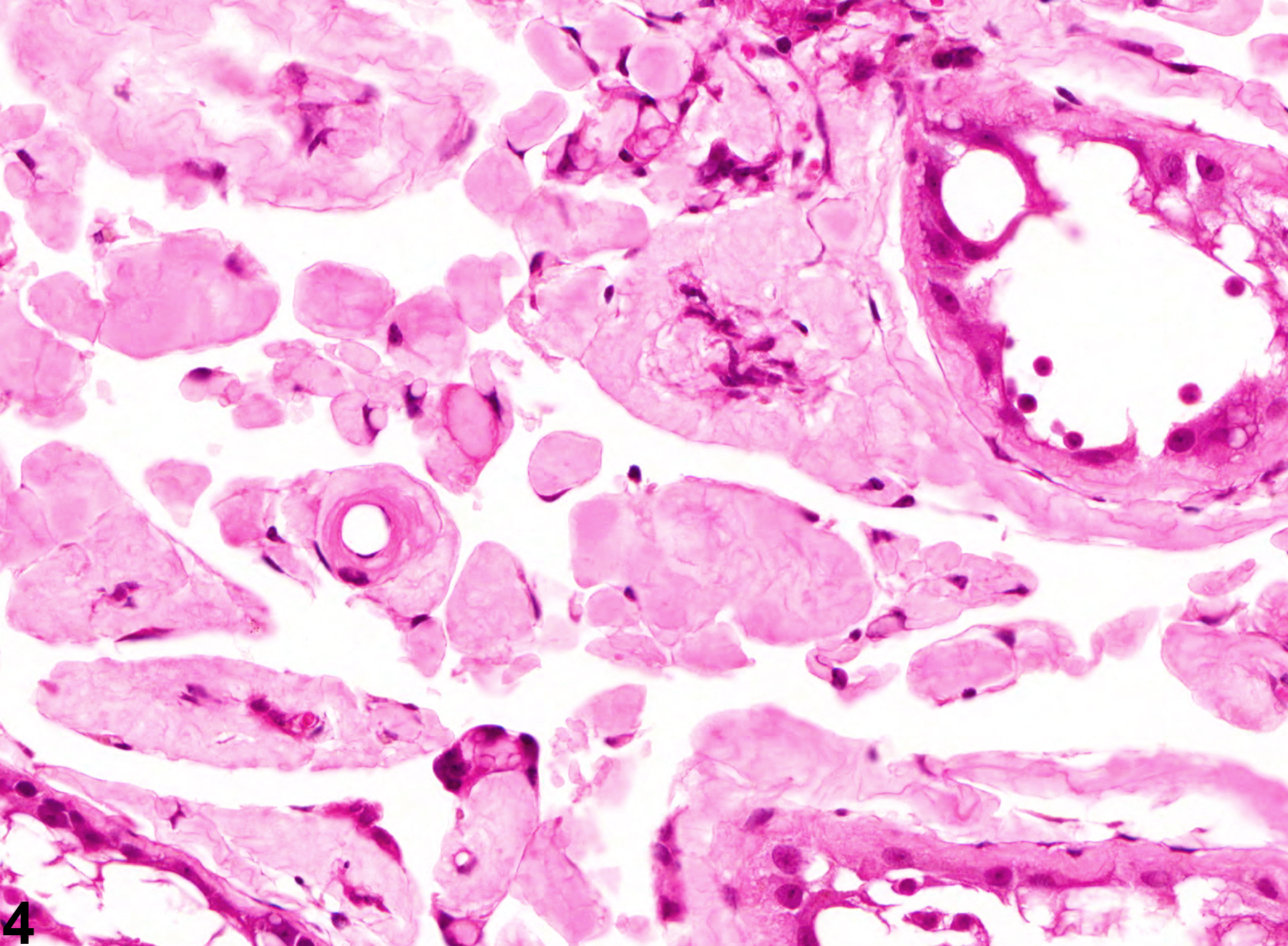
Testis - Amyloid in a male Swiss CD-1 mouse from a chronic study. Higher magnification of Figure 3 showing extensive amyloid deposition.