U.S. flag

An official website of the United States government

Dot gov

The .gov means it's official.
Federal government websites often end in .gov or .mil. Before sharing sensitive information, make sure you're on a federal government site.

Https

The site is secure.
The https:// ensures that you are connecting to the official website and that any information you provide is encrypted and transmitted securely.

Skip to Main Navigation
Skip to Page Content
Skip to Atlas Navigation

Testis, Germ Cell - Exfoliation

Image of germ cell exfoliation in the testis from a male Harlan Sprague-Dawley rat
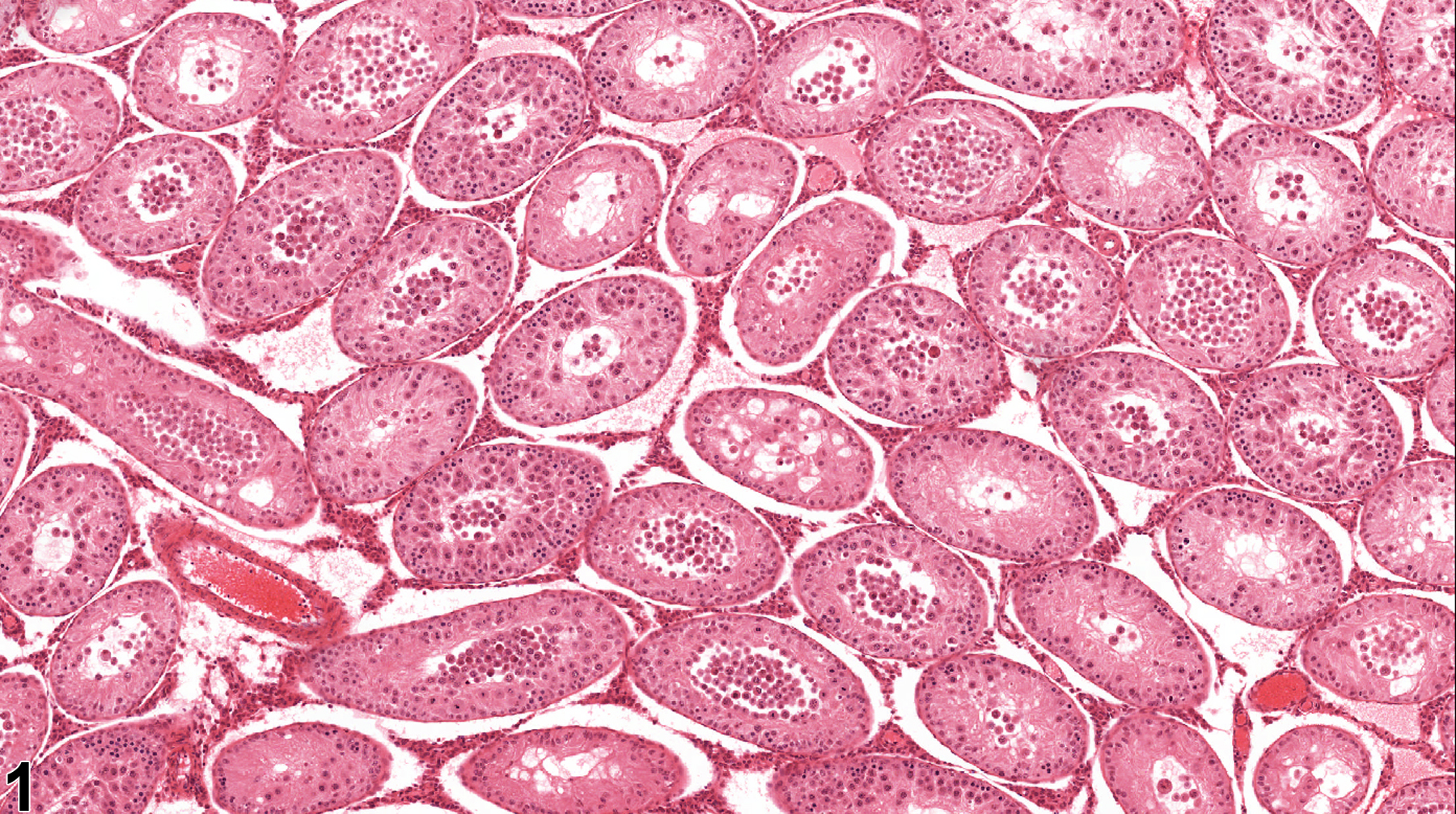
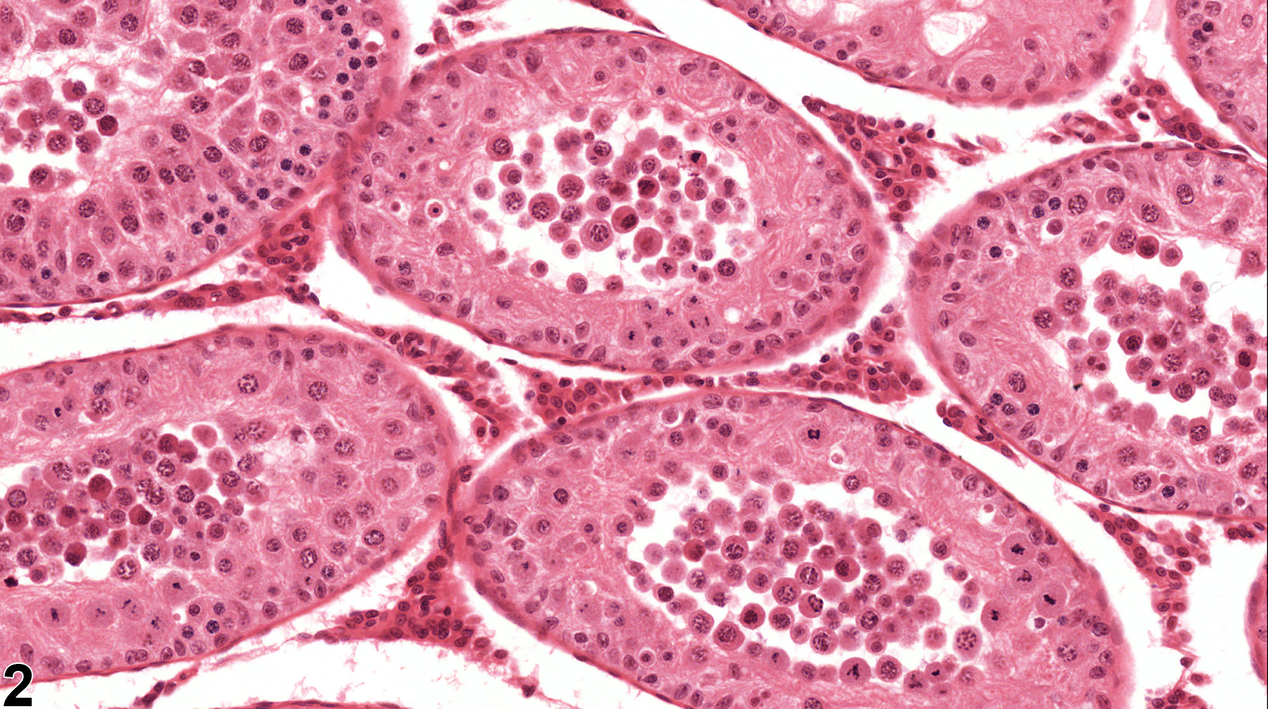
Testis, Germ cell - Exfoliation in a male Harlan Sprague-Dawley rat. Exfoliated germ cells are present in seminiferous tubule lumens. (Photograph courtesy of Dr. D. Creasy.)
Figure 1 of 2
Image of germ cell exfoliation in the testis from a male Harlan Sprague-Dawley rat
Testis, Germ cell - Exfoliation in a male Harlan Sprague-Dawley rat. Higher magnification of Figure 1 showing germ cells in seminiferous tubule lumens. (Photograph courtesy of Dr. D. Creasy.)
Figure 2 of 2
next arrow

comment:

Germ cells may undergo degeneration and/or apoptosis within the seminiferous epithelium, or they may lose contact with the surrounding Sertoli cell cytoplasmic processes and be shed into the tubular lumen ( Figure 1image opens in a pop-up window and Figure 2image opens in a pop-up window ). Often the exfoliated germ cells retain relatively normal morphology. The cells will rapidly be transported into the epididymis and will be present in the ductular lumens. The finding should not be confused with artifactual sloughing of germ cells caused by squeezing of the testis during necropsy dissection. In the case of sloughing due to necropsy trauma, germ cells will not be seen in the epididymis. Germ cell exfoliation has been described as a predominant finding following administration of phthalate esters and microtubule inhibitors such as colchicine and the fungicide carbendazim.

recommendation:

The term "germ cell exfoliation" should be reserved for those situations where there is extensive shedding of germ cells into the lumen as a predominant finding. In those situations, germ cell exfoliation should be diagnosed and graded and should be discussed in the pathology narrative if the incidence and/or severity appears to be related to chemical administration. Bilateral involvement should be diagnosed when present. If there is significant concurrent germ cell degeneration, that should also be diagnosed and given a severity grade.

references:

Correa LM, Nakai M, Strandgaard CS, Hess RA, Miller MG. 2002. Microtubules of the mouse testis exhibit differential sensitivity to the microtubule disruptors carbendazim and colchicine. Toxicol Sci 69:175-182.
Abstract: http://www.ncbi.nlm.nih.gov/pubmed/12215672

Creasy DM. 2001. Pathogenesis of male reproductive toxicity. Toxicol Pathol 29:64-76.
Full Text: http://tpx.sagepub.com/content/29/1/64.full.pdf

Creasy D, Bube A, de Rijk E, Kandori H, Kuwahara M, Masson R, Nolte T, Reams R, Regan K, Rehm S, Rogerson P, Whitney K. (2012). Proliferative and nonproliferative lesions of the rat and mouse male reproductive system. Toxicol Pathol 40:40S-121S.
Abstract: https://doi.org/10.1177/0192623312454337

Nakai M, Hess RA. 1994. Morphological changes in the rat Sertoli cell induced by the microtubule poison carbendazim. Tissue Cell 26:917-127.
Abstract: http://www.ncbi.nlm.nih.gov/pubmed/7886678

Russell LD, Malone JP, MacCurdy DS. 1981. Effect of the microtubule disrupting agents, colchicine and vinblastine, on seminiferous tubule structure in the rat. Tissue Cell 13:349-367.
Abstract: http://www.ncbi.nlm.nih.gov/pubmed/7314074