

Reproductive System, Male
Testis, Interstitial Cell - Syncytial Cells
Narrative
Comment:
The syncytial cell formation is unusual and has not been described in the literature. The interstitial syncytial cells may represent Leydig cells or macrophages. These can be distinguished using a Periodic acid-Schiff stain, which stains macrophages but not Leydig cells. The pathogenesis and significance of this lesion are unknown.
Recommendations:
Interstitial syncytial cells should be diagnosed and graded when present and should be discussed in the pathology narrative if the incidence and/or severity appears to be related to chemical administration. Diagnosis should indicate bilaterality when present.
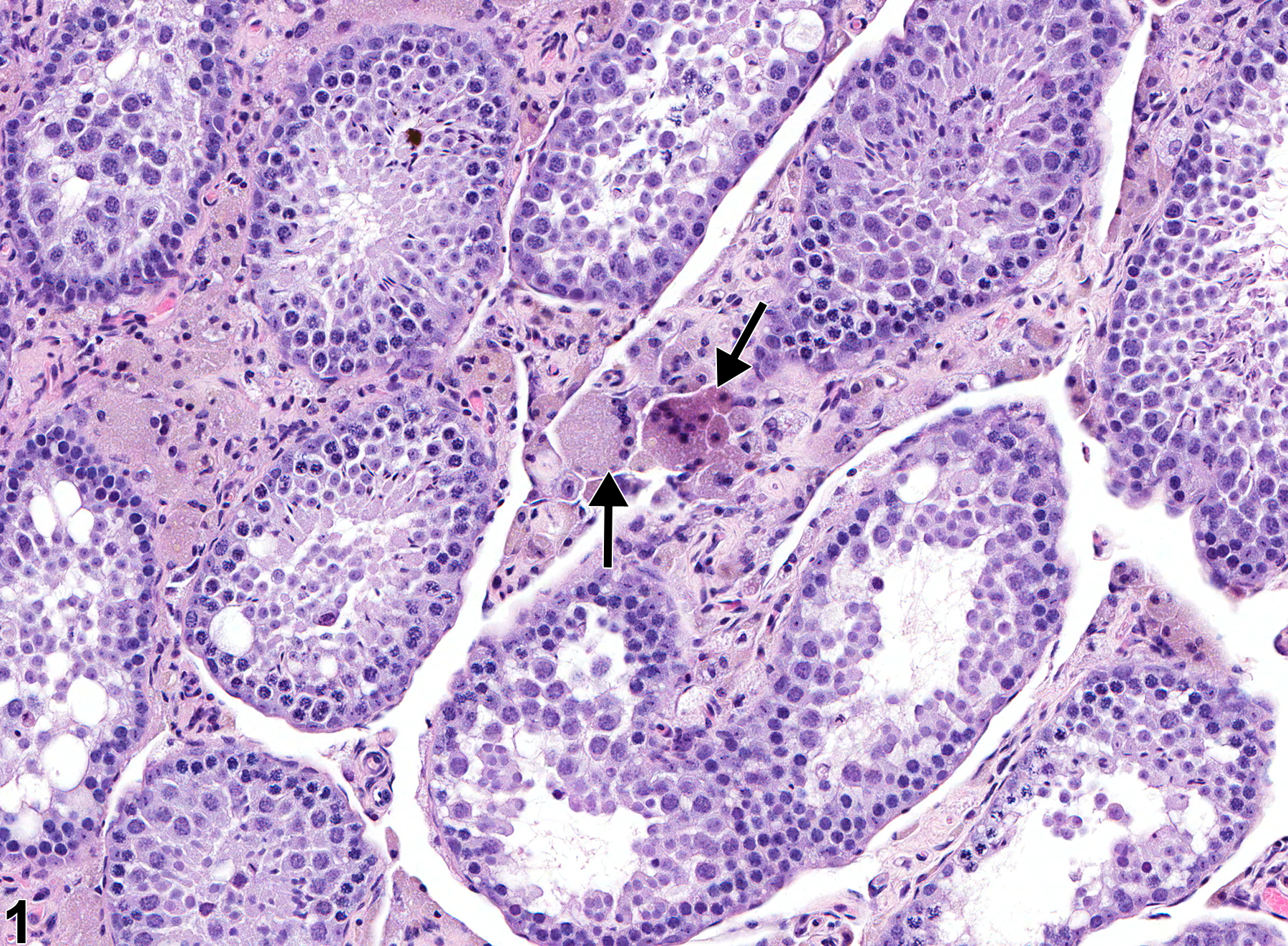
Testis, Interstitial cell - Vacuolation in a male FVB/N transgenic mouse from a chronic study. There are interstitial syncytial cells (arrows).
All Images

Testis, Interstitial cell - Vacuolation in a male FVB/N transgenic mouse from a chronic study. There are interstitial syncytial cells (arrows).
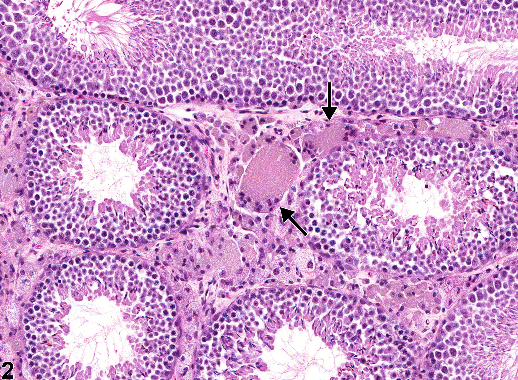
Testis, Interstitial cell - Vacuolation in a male FVB/N transgenic mouse from a chronic study. There are interstitial syncytial cells (arrows) accompanied by vacuolation of Leydig cells.