

Reproductive System, Male
Testis, Interstitial Cell - Vacuolation
Narrative
Comment:
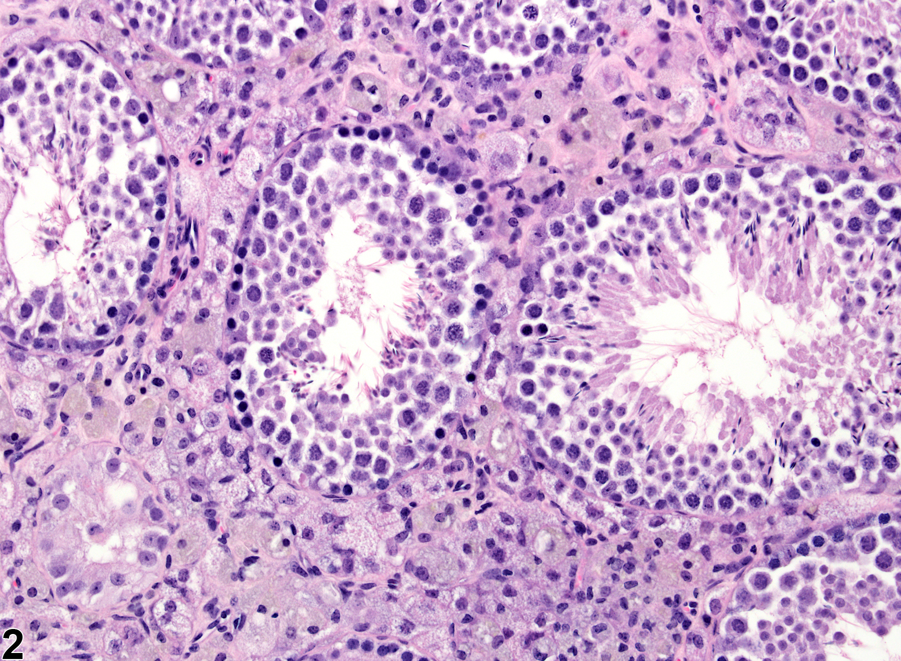
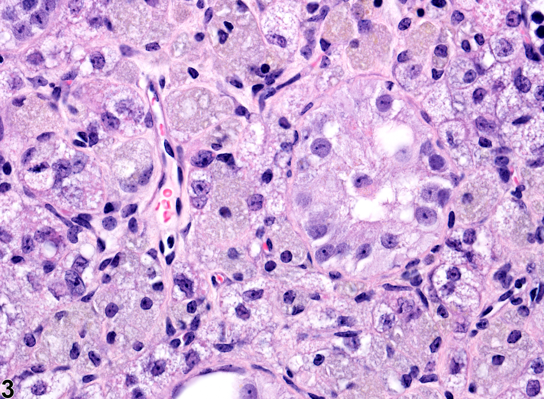
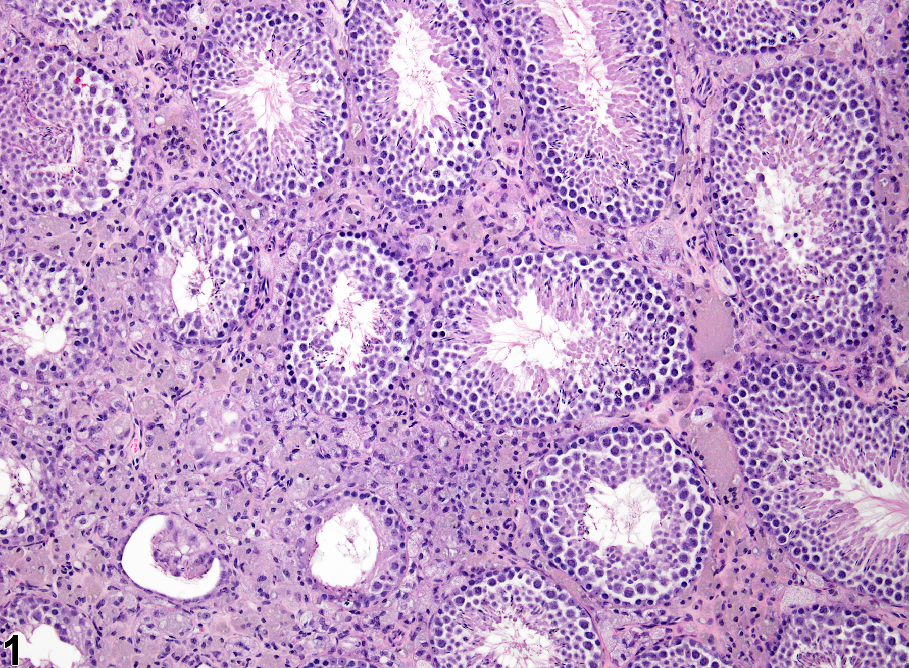
Cytoplasmic vacuolation of interstitial cells can be seen as a chemically induced change and most likely represents a disturbance in steroidogenesis. It generally consists of enlarged, pale-staining interstitial cells with foamy vacuolation of the cytoplasm (Figure 1, Figure 2, and Figure 3). Syncytial giant cell formation is also present in the interstitial compartment of the testes of these mice with vacuolated interstitial cells. The transgenic mice in Figure 1, Figure 2, and Figure 3 were treated with ethinyl estradiol. Testicular macrophages can also become vacuolated, independent of interstitial cells. The vacuolated cells may represent interstitial cells or macrophages. These can be distinguished using a Periodic acid-Schiff stain, which stains macrophages but not interstitial cells. Note that normal interstitial cells of mice are generally larger and have a more vacuolated appearance than those of rats.
Recommendations:
Interstitial cell vacuolation should be diagnosed and graded and should be discussed in the pathology narrative if the incidence and/or severity appears to be related to chemical administration. Diagnosis should indicate bilaterality when present.
Testis, Interstitial cell - Vacuolation in a male FVB/N transgenic mouse from a chronic study. The interstitial cells contain variably sized vacuoles.
All Images

Testis, Interstitial cell - Vacuolation in a male FVB/N transgenic mouse from a chronic study. The interstitial cells contain variably sized vacuoles.

Testis, Interstitial cell - Vacuolation in a male FVB/N transgenic mouse from a chronic study. This higher magnification of Figure 1 shows the vacuolation of interstitial cells.

Testis, Interstitial cell - Vacuolation in a male FVB/N transgenic mouse from a chronic study. This higher magnification image of Figure 2 shows the vacuoles in detail, as well as some golden brown pigment.